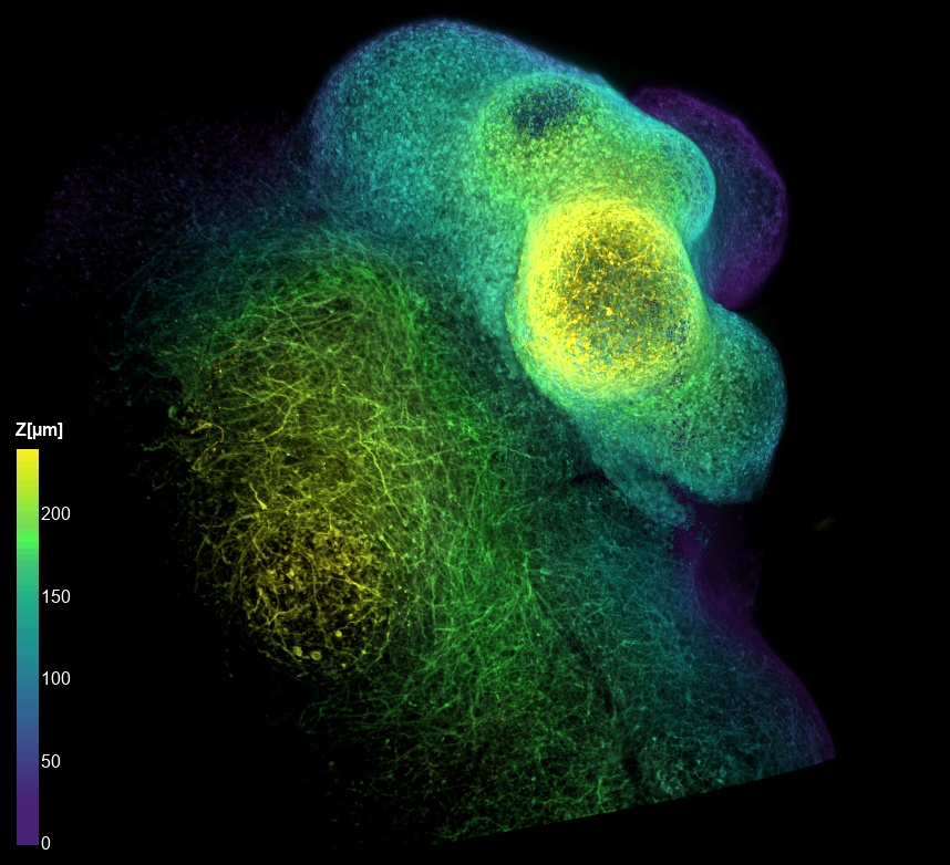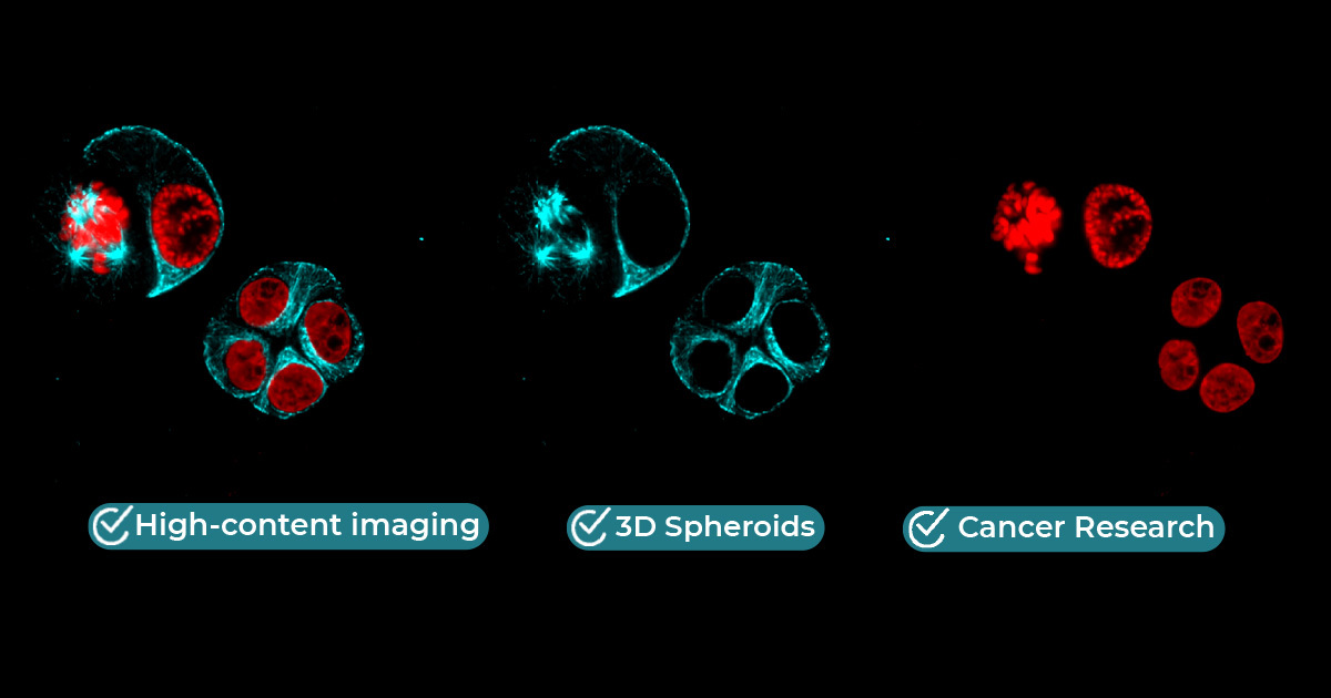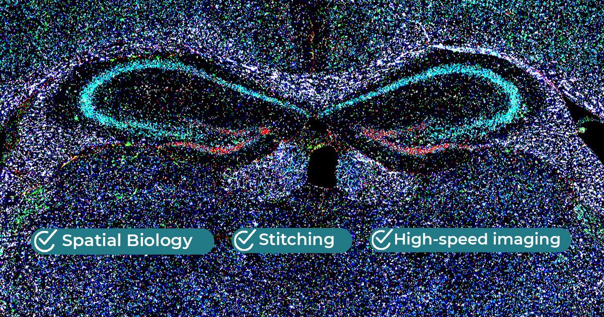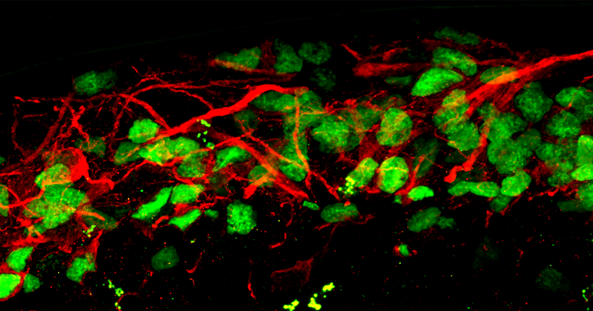
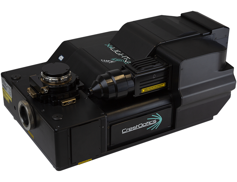
"Compared to other systems, the X-Light V3 spinning disk is exceptionally fast, making it highly effective for volumetric imaging of large 3D tissues. By utilizing its fast time-lapse capabilities, we achieve the crucial temporal resolution needed for highly dynamic functional assays, all while maintaining an excellent field of view."
“The X-Light V3’s high-speed, low-phototoxic imaging allows us to visualize dynamic processes and advanced 3D models in real time. This provides unique insights into complex mechanisms which are essential for identifying novel therapeutic strategies.”
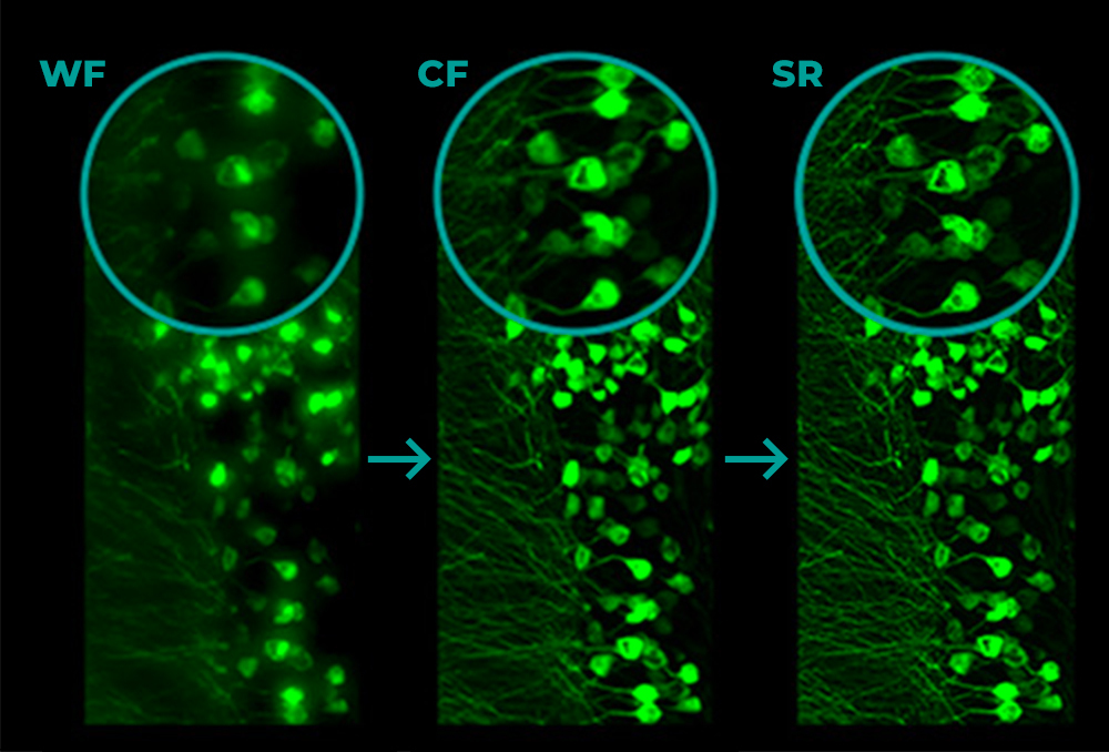
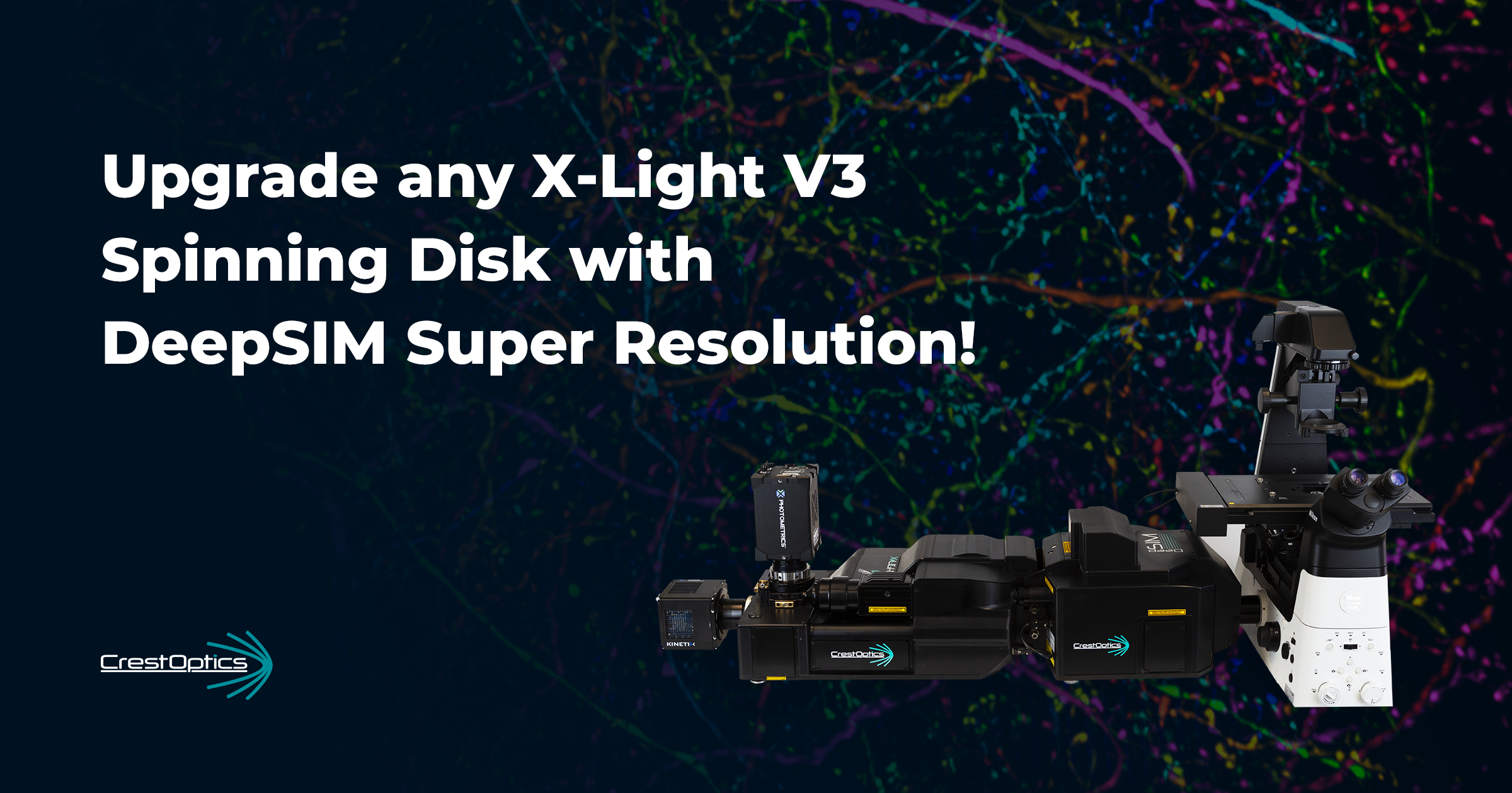
"The versatility of combining Widefield, Spinning Disk, and Super-Resolution in a single system allows us to address a wide variety of research needs without the need for multiple instruments."

“The microscope equipped with X-Light V3 represents a very versatile platform, and it is adaptable with excellent results to the multiple applications I work on”
“The unique features of the CrestOptics X-Light V3 Spinning Disk confocal unit, such as the easily interchangeable disk unit combined with the large FOV, illumination shaping and uniformity are the key points which convinced us to adopt the system.”
Dr. Gaia Pigino Human Technopole (Milan)
“Facility users appreciate a lot the easy switch from Widefield to Spinning disk modality, giving them the possibility to select the proper imaging according to their application and experimental needs.”

Dr. Gabriela ImrehKarolinska Institutet (Stockholm)
“The main goal we wanted to reach was to have a high-throughput spinning disk system and we largely met our needs with the CrestOptics X-Light V3.”