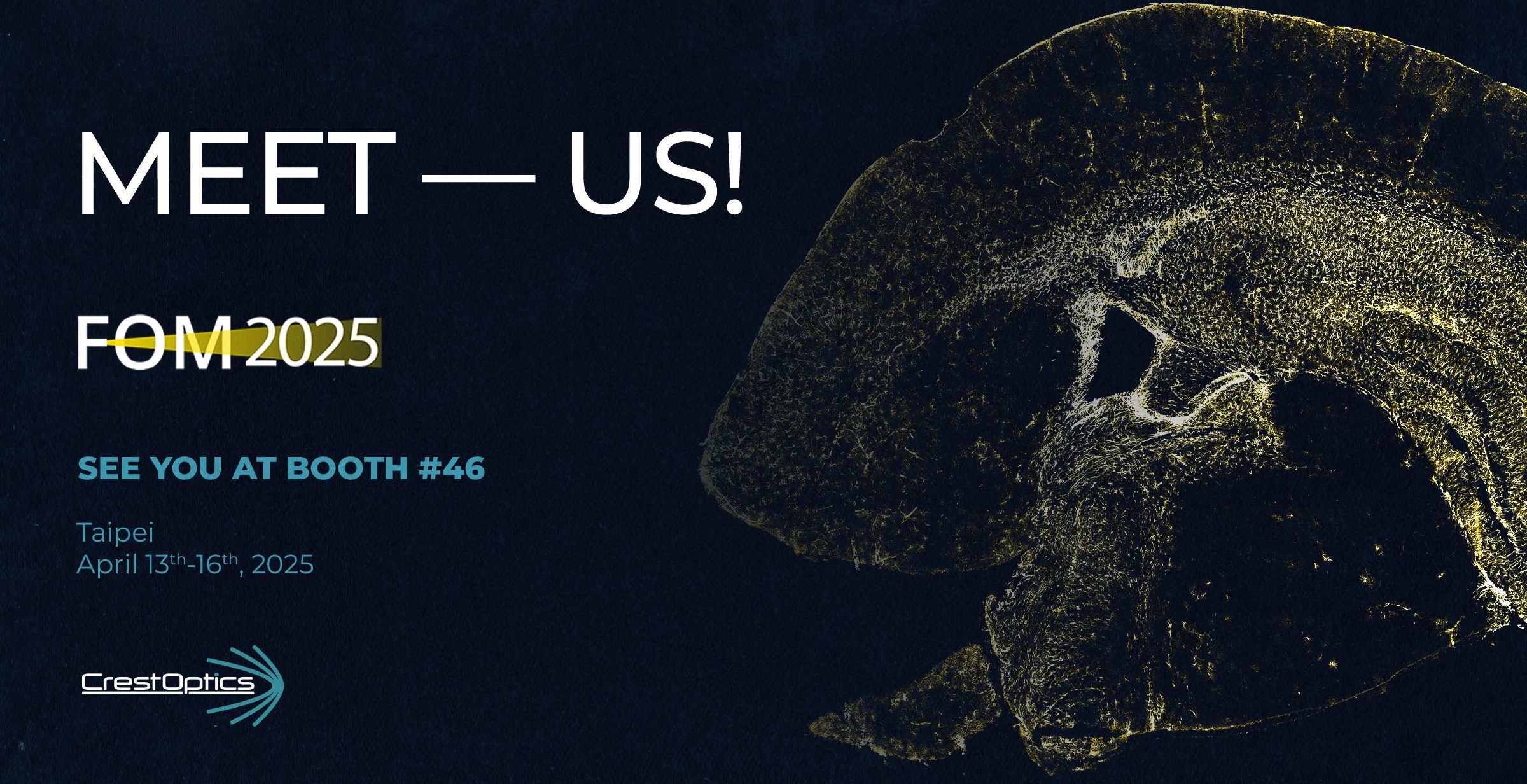
April 13-16, 2025
Join Us at Focus on Microscopy 2025
CrestOptics is excited to participate in FOM 2025 (Focus on Microscopy) in Taipei, Taiwan, 13-16th April 2025, a globally recognized conference that brings together experts and professionals in the field of microscopy. This event serves as a key platform for showcasing the latest innovations and advancements in imaging technology.
Explore Our Microscopy Solutions
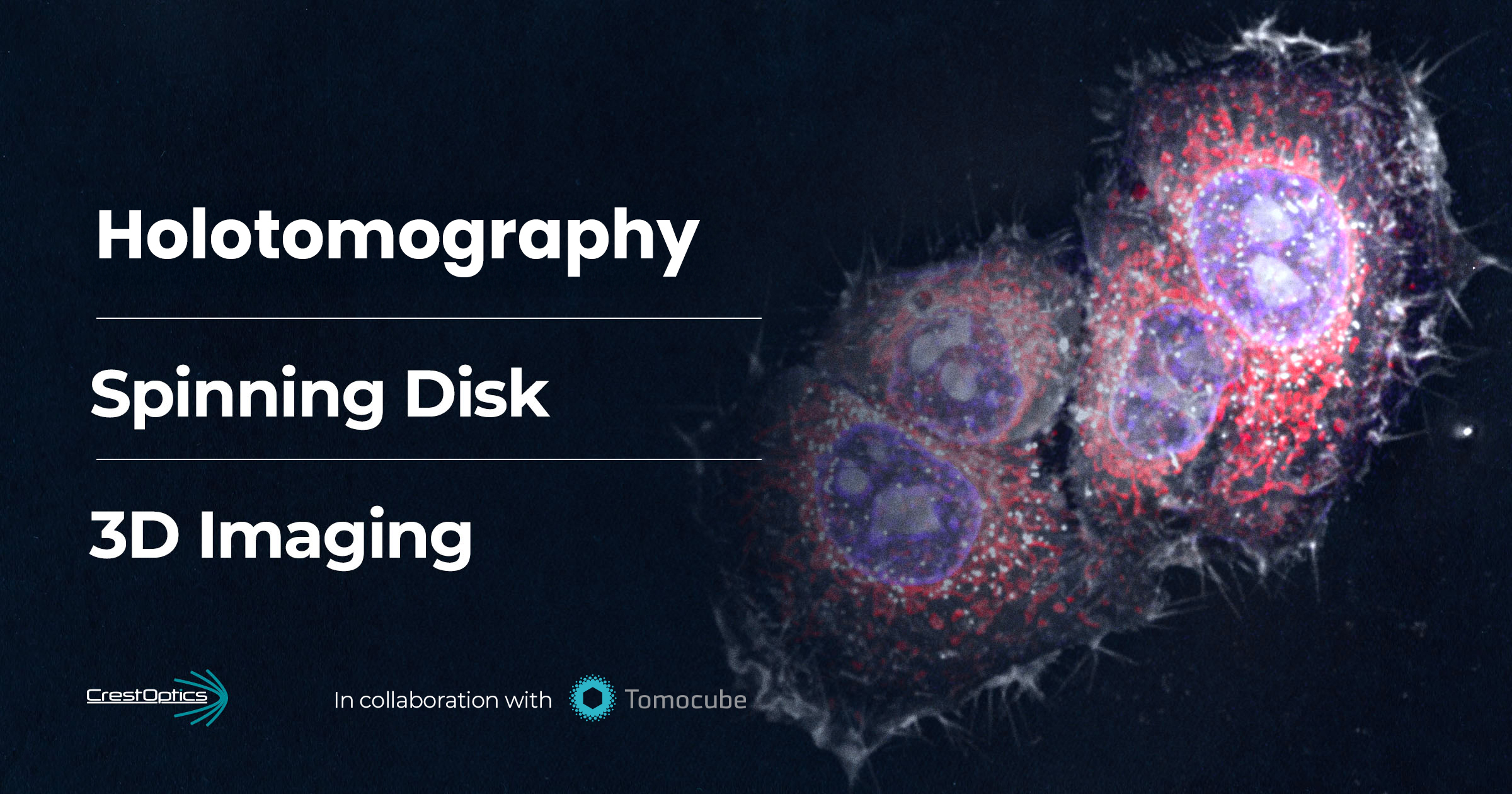
At booth #46, we will showcase our high-performance and accessible microscopy solutions: X-Light V3 and CICERO, specifically designed to advance cutting-edge research in life sciences. Our solutions offer:
- Seamless Integration – Easily adaptable to existing workflows
- Maximum Configuration Flexibility – Enabling a wide range of imaging applications
- State-of-the-Art Imaging Technologies – Delivering high-performance results
If you are attending FOM, don’t miss our workshops!
Throughout the conference, we will be hosting three workshops on X-Light V3 spinning-disk and DeepSIM super-resolution technologies, followed by hands-on microscopy sessions. The total duration of each workshop is approximately 2 hours (30 minutes of presentation followed by a practical demonstration).
Below, you will find the details for each of the three workshops, click the registration button to secure your spot!
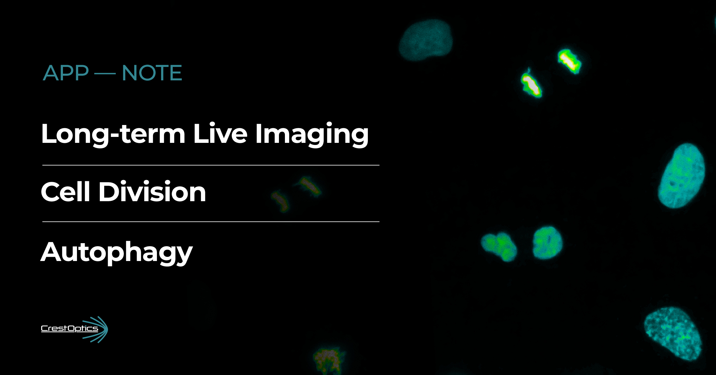
1) Imaging Across Scales:
Sample Flexibility and Versatile Applications
| Sunday – April 13rd | 10 AM session and 4 PM session
Read the workshop abstract
Abstract:
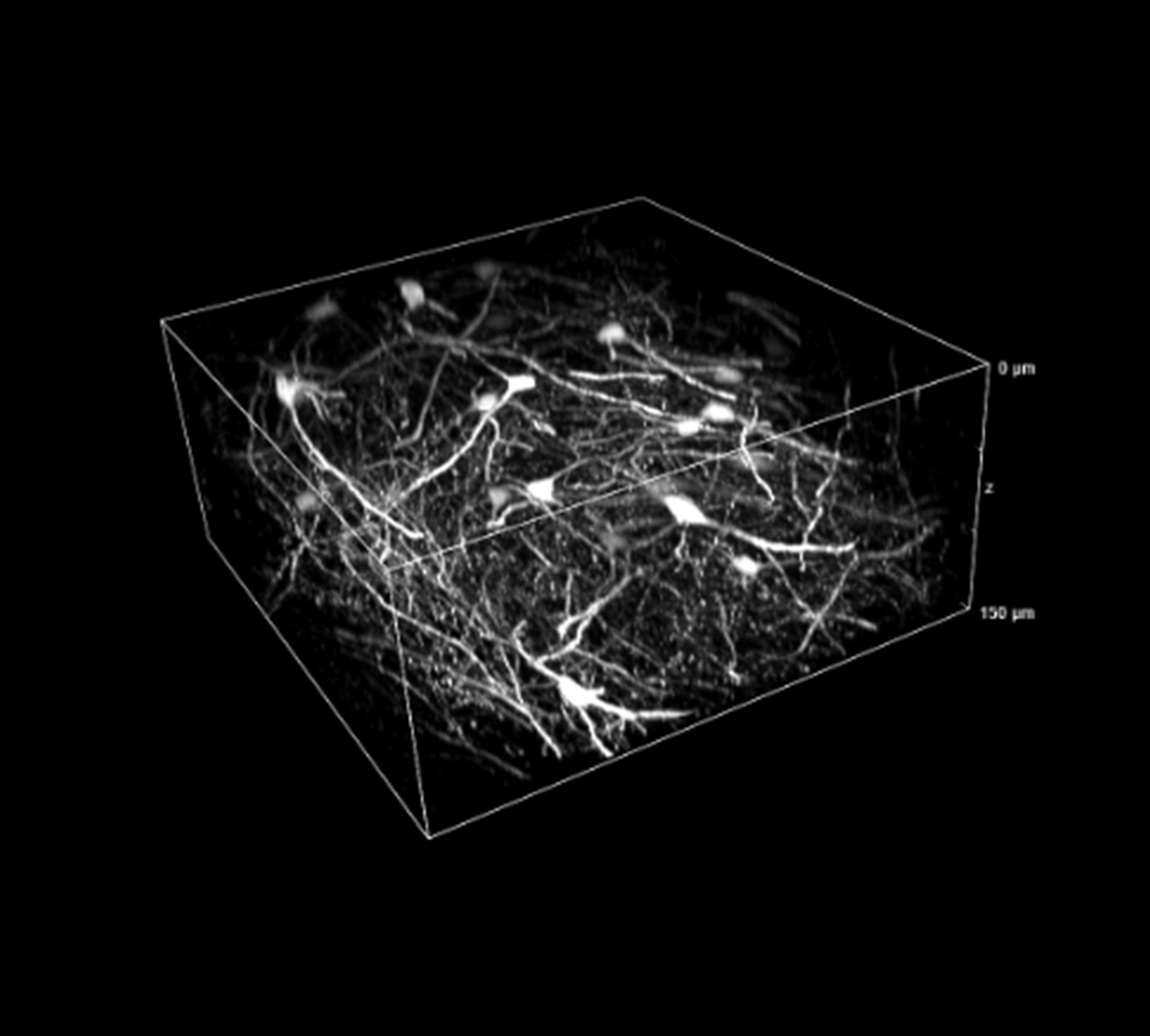
Biological imaging requires versatile tools capable of adapting to a wide range of samples, from thick tissues to three-dimensional models such as organoids and spheroids, down to single cells and subcellular structures. The combination of the X-Light V3 system, for fast and high-contrast confocal imaging, and DeepSIM, for super-resolution, provides a scalable approach to analyzing biological structures across different spatial scales. This allows researchers to capture both the global context of a tissue and ultrastructural details with precision, enabling advanced studies in cell biology, neuroscience, and oncology. The high compatibility with samples of various thicknesses and compositions makes this platform ideal for applications ranging from in vivo imaging to high-resolution analysis of fixed specimens.
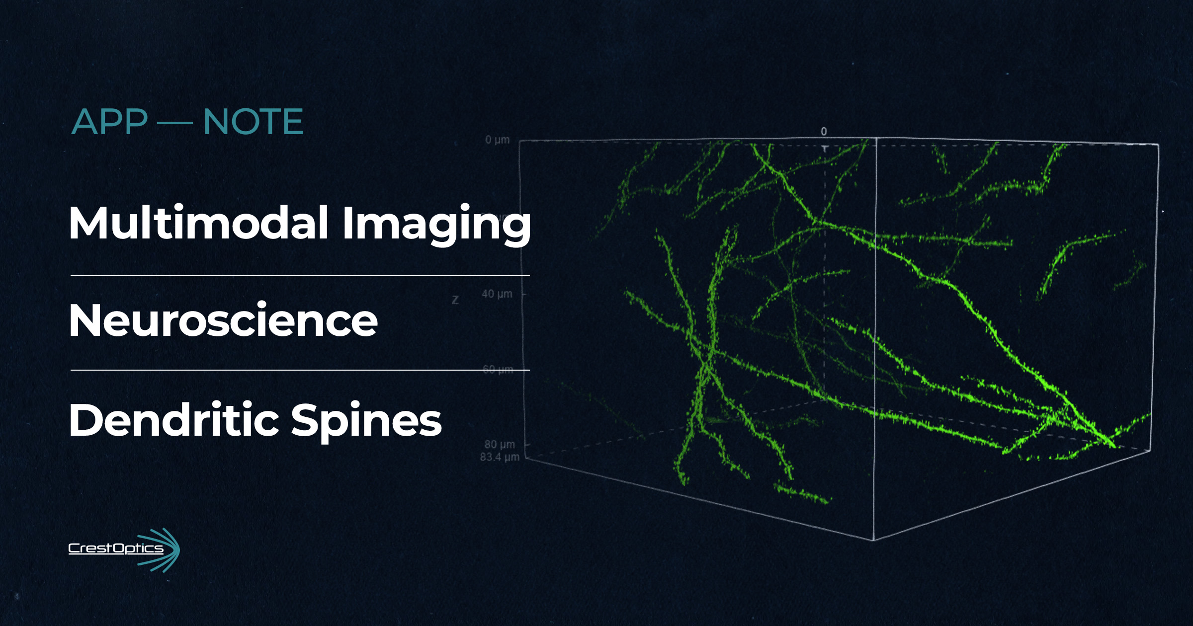
2) Multimodal Imaging:
Integrating Three Modalities for Comprehensive Analysis
| Monday – April 14th | 10.30 AM session and 3 PM session
Read the workshop abstract
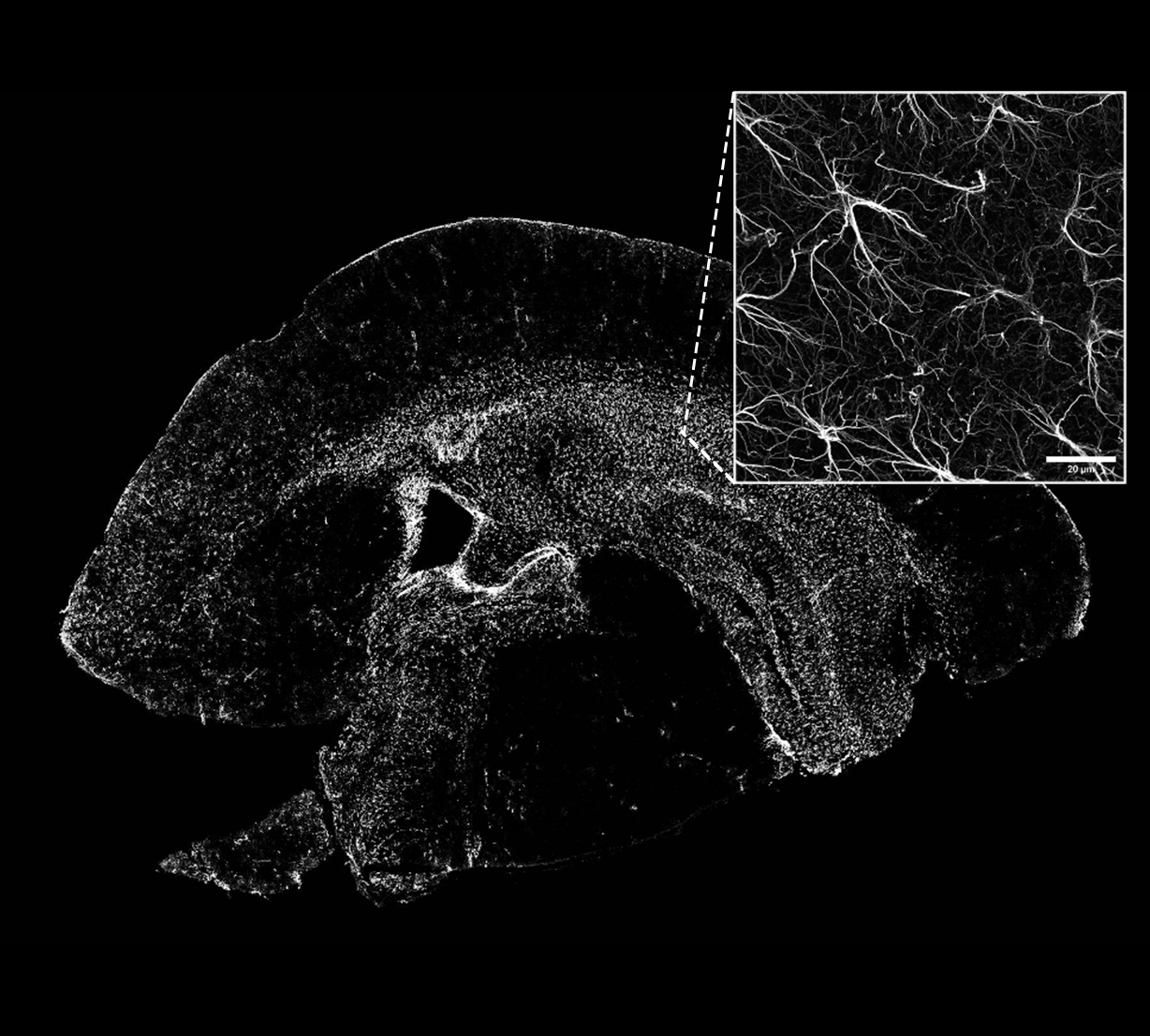
Integrating multiple imaging modalities within a single platform enables the acquisition of complementary information, combining speed, sensitivity, and resolution. The Widefield + Confocal Spinning Disk (X-Light V3) + Super-Resolution (DeepSIM) configuration allows seamless transitions between imaging modes, optimizing the balance between field of view, contrast, and spatial resolution. Widefield imaging is ideal for rapid screening and large-area visualization, while confocal mode enables optical sectioning for deeper sample analysis. Finally, super-resolution imaging enhances the visualization of subcellular details, essential for studying molecular interactions and structural organization. This multimodal approach represents a powerful solution for research in cell biology, neuroscience, and biomedical sciences, providing quantitative data across different spatial scales with an optimized workflow.
3) Spatial-Omics and Super-Resolution:
Enhancing Spot Detection Efficiency
| Tuesday – April 15th | 10.30 AM session and 3 PM session
Read the workshop abstract
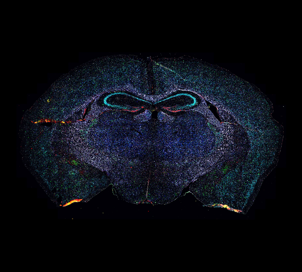
Spatial-omics technologies, including spatial transcriptomics and proteomics imaging, are revolutionizing molecular biology by enabling the precise mapping of gene and protein expression directly within tissue samples. However, the detection of molecular spots often faces limitations due to the optical resolution of conventional techniques, which can hinder accurate data interpretation. The DeepSIM system addresses these challenges by surpassing the diffraction limit and achieving lateral resolution as fine as ~100 nm, allowing for more precise and quantitative detection of fluorescent signals in spatial-omics analyses. This improved resolution enhances sensitivity, enabling the detection of low-abundance transcripts and proteins, thus boosting the accuracy of spatial datasets. Moreover, we investigate the impact of structured-illumination microscopy (SIM) in combination with both low- and high-magnification objectives for the localization of individual fluorescent amplicon spots. The widely used 20× 0.8 NA objective, when paired with super-resolution or spinning disk confocal imaging, provides sufficient detail for identifying low-expression genes. The DeepSIM system also excels in resolving high-density regions and distinguishing closely packed spots within specific areas. With its compatibility with standard fluorophores and seamless integration into existing workflows, the DeepSIM is an invaluable tool for advanced imaging applications in molecular biology and personalized medicine.
Let’s connect!
Attendees will have the opportunity to engage with our experts to learn how CrestOptics technologies can enhance their research. FOM 2025 is not only a place to explore cutting-edge technology but also an opportunity to connect with the people behind CrestOptics. Visit our booth #46 to discuss your research needs and discover how our microscopy solutions can accelerate your experiments.