In a recent study published in Cell Death and Disease (Brighi et al., 2021), resulting from the collaboration between Prof. Silvia Di Angelantonio and Prof. Alessandro Rosa at the Joint Lab between Center for Life Nano & Neuro-Science and Sapienza University of Rome (CL2NS@Sapienza – Italian Institute of Technology), novel human 2D and 3D brain models have been generated to reproduce some of the neurobiological phenotypes associated with Fragile X Syndrome (FXS). In this work, the authors demonstrate the possibility to study FXS using human cerebral organoids suggesting, for the first time, the use of this experimental platform to model FXS in a human genetic context.
FXS is an inherited neurodevelopmental disorder caused by epigenetic silencing in the FMR1 gene and the consequent loss of the fragile X mental retardation protein (FMRP).
In FXS patients, alterations in dendritic spine morphology, synaptogenesis and connectivity in the developing brain lead to cognitive impairment, defective communication, hyperactivity and anxiety.
Over the years, several animal models have been used for the investigation of FXS and its mechanisms. Nevertheless, despite FXS animal models, especially the Fmr1 KO mouse, represent a fundamental resource in understanding the different mechanisms inherent in this pathology, physiological and evolutionary discrepancies have hindered the translation of these results from animals to humans. For these reasons, the authors produced an in vitro FXS model system based on isogenic mutant (FMRP-KO) and control (FMRP-WT) human induced pluripotent stem cells (hiPSCs) lines, demonstrating that advances in disease-relevant hiPSC generation, and modification by genome editing, provide novel possibilities for FXS disease modelling.
In this work, these lines were first differentiated to cortical neurons using 2D culture conditions and, as cortical hyperexcitability represents one of the hallmarks of FXS, the authors characterized the development of the glutamatergic and GABAergic systems through confocal analysis of immunofluorescence signals. For this purpose, in Brighi et al. a CrestOptics X-Light V3 spinning disk was used to acquire images to investigate the presence and the proper formation of glutamatergic and GABAergic synaptic components. Therefore, a side-by-side comparison of FMRP-WT and FMRP-KO cultures revealed that FMRP-KO neurons at day 54 displayed higher number of pre- (VGLUT1) and post- (PSD95) synaptic glutamatergic components demonstrating a temporary increase of excitatory glutamatergic synaptic markers during in vitro maturation (see Figure 3 and Figure 4 of Brighi et al. for the images and the method section “immunostaining and image acquisition and analysis of 2D cultures” for analysis details).
Further analyses on FMRP-KO 2D cultures have highlighted an altered neuronal and glial gene expression and proliferation, an increased network activity and a pronounced hyperexcitability, demonstrating how these 2D in vitro models are able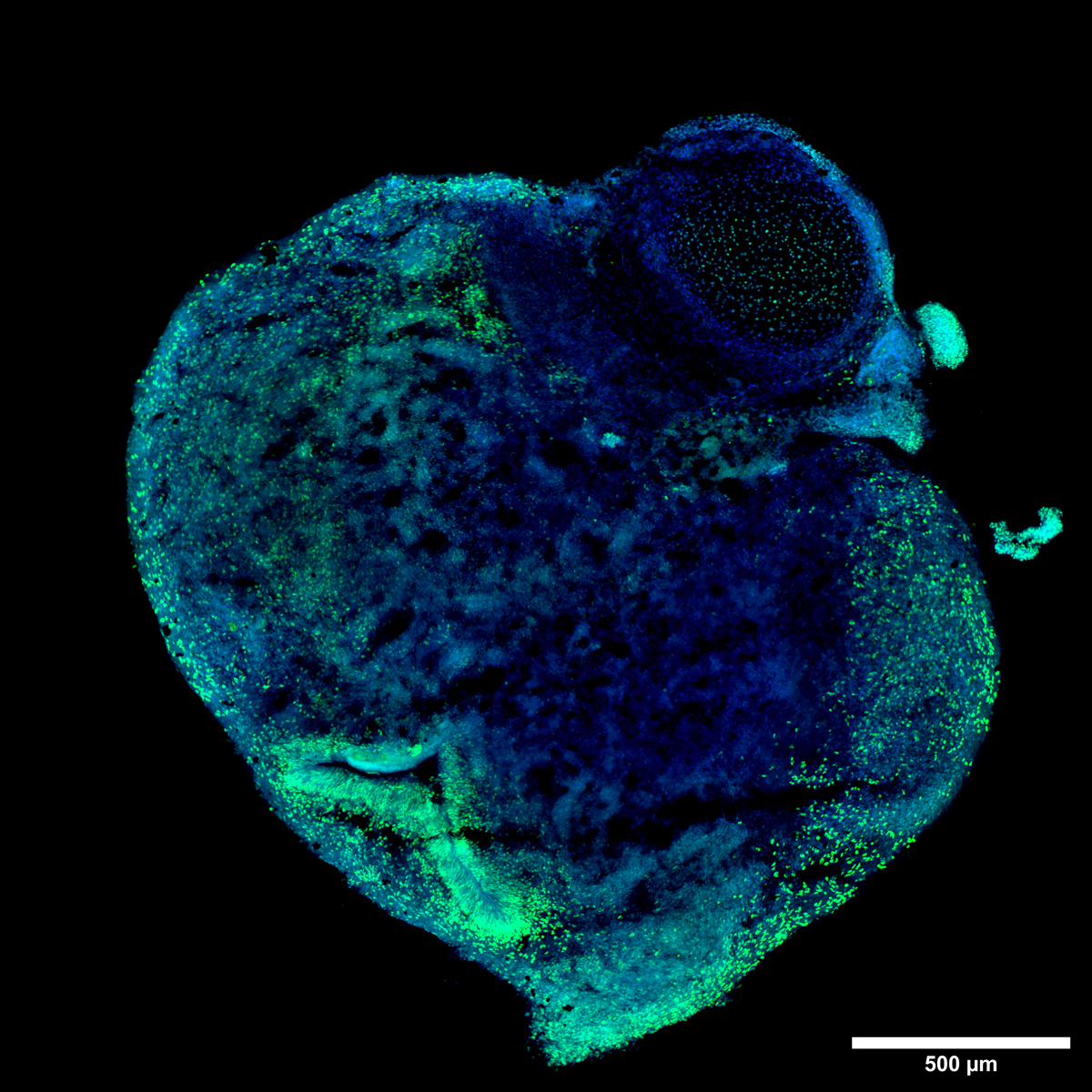
Figure A: Large acquisition of a day 50 human cerebral organoid displaying a clear organization of SOX2+ neural progenitor cells (green) forming organized structures resembling the ventricular and subventricular zone. Nuclei were stained with DAPI (blue). Scale bar: 500 um. This image was acquired with a CrestOptics X-Light V3 spinning disk.
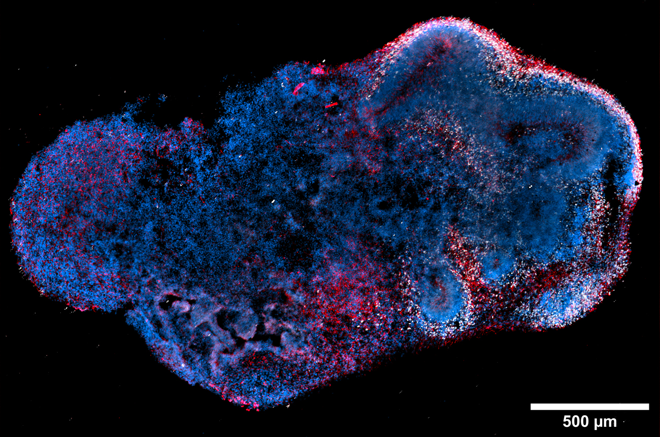
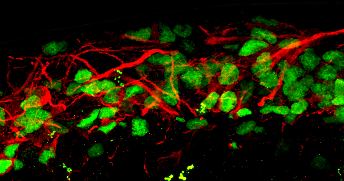
Figure B: Large image of a day 50 human cerebral organoid showing CTIP2-positive deep layer cortical neurons in white, and pan-neuronal MAP2 marker signal in red. Nuclei were stained with DAPI (blue). Scale bar: 500 um. This image was acquired with a CrestOptics X-Light V3 spinning disk.
In the same way, as represented in Figure B and similarly to what shown in Brighi et al. paper, it is possible to easily spot the presence of cortical deep-layer neurons (CTIP2+ in white and MAP2+ in red) that, differentiating towards the periphery, shape the typical laminar cortical structure.
Once the area of interest is identified, it is easier to investigate the three-dimensional biological complexity of the cortical region with higher magnification, deepening the cortical neurons layer organization (Figure C).
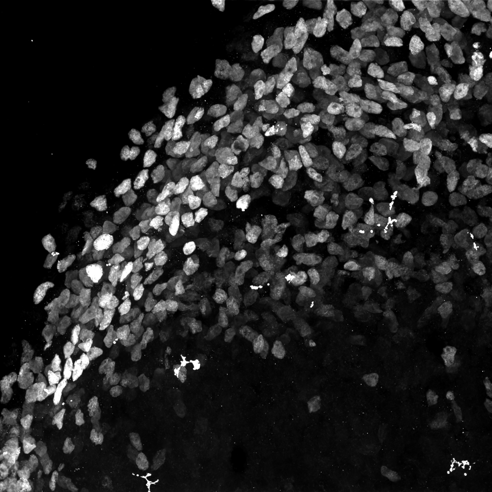
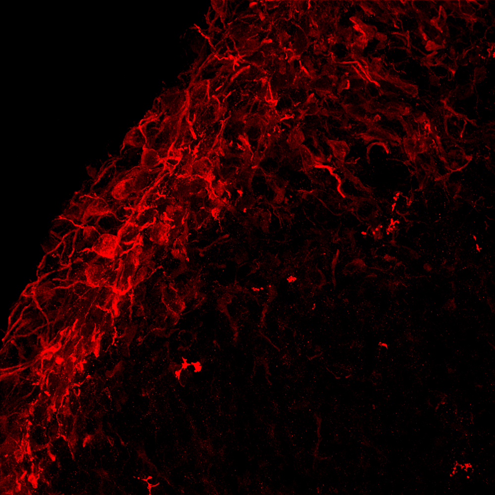
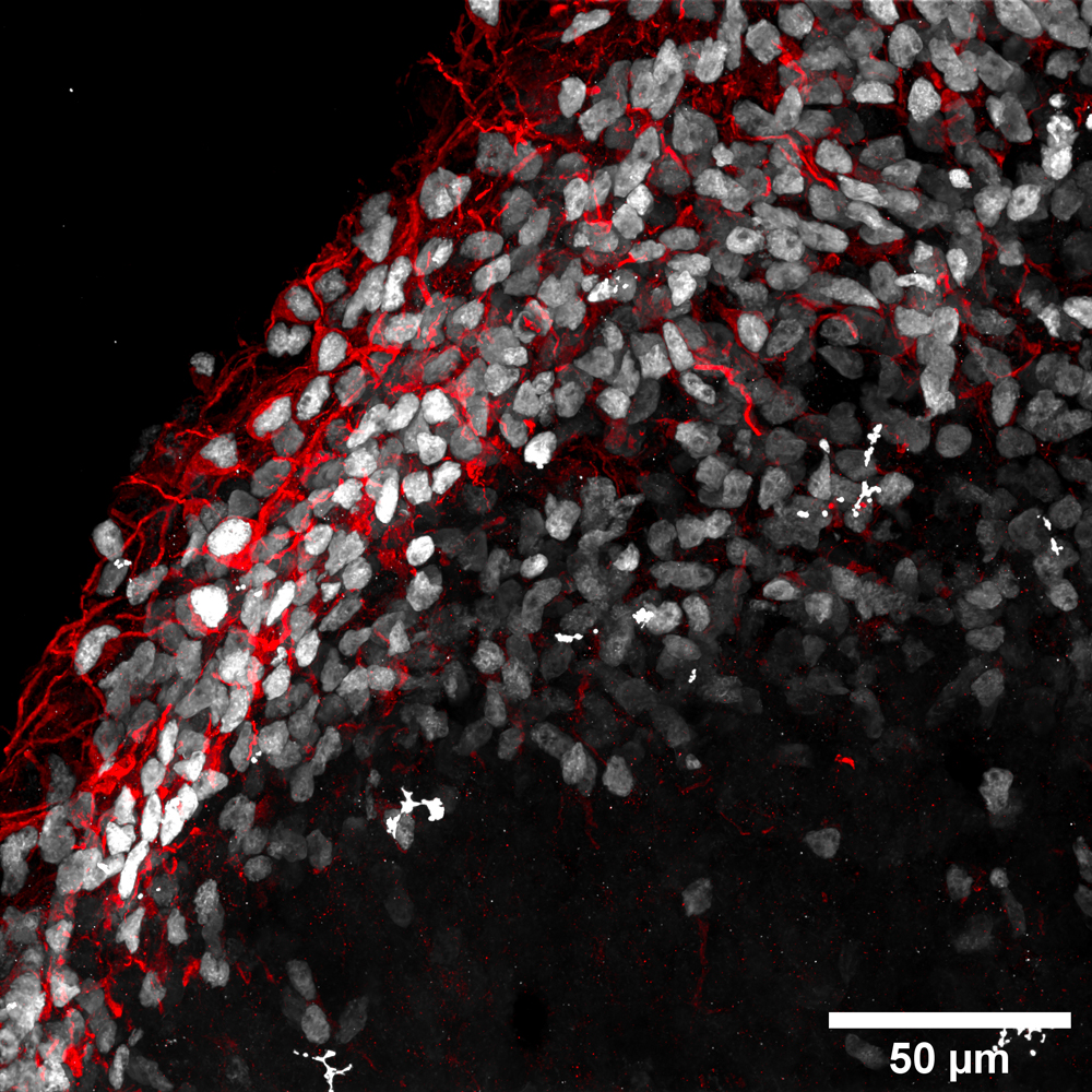
Figure C: Maximum intensity projection of cortical neurons positive for CTIP2 (white) and pan-neuronal MAP2 marker (red). Scale bar: 50 um. This image was acquired with a CrestOptics X-Light V3 spinning disk.
In conclusion, with CrestOptics X-Light V3 spinning disk it is possible to obtain in few minutes an image that represents a sample as complex as that of the cerebral organoids in its entirety, also using multiple wavelengths during high-sectioning of its entire thickness. At the same time, it is possible to go into more detail with higher magnifications to appreciate the three-dimensional architecture that the cells assume only in some portions of the sample, obtaining a very high-resolution image and, at the same time, avoiding photobleaching issues thanks to the extreme speed of this spinning disk system.
MICROSCOPY METHODS
For 2D images acquisitions and analysis details see the method section “immunostaining and image acquisition and analysis of 2D cultures” of Brighi et al.
The acquisition of cerebral organoids images shown here was performed through a Nikon Eclipse Ti2 microscope equipped with X-Light V3 spinning disk (CrestOptics), Celesta laser source (Lumencore) and Prime BSI Scientific CMOS (sCMOS) camera with 6.5 um pixels (Photometrics). The images were acquired by using NIS-Elements Microscope Imaging software version 5.30.02 (Nikon).
Figure A represents an intensity projection (MIP) of a day 50 human cerebral organoid and was acquired with a CFI Plan Apo Lambda 20X air objective (NA 0.75, WD 1) in a stack with z-step of 0.9 um and with 23 um of Z range.
Figure B shows a MIP of a day 50 human cerebral organoid acquired with a CFI Plan Apo Lambda 20X air objective (NA 0.75, WD 1) in a stack with z-step of 2 um and with 20 um of Z range.
Figure C images illustrate MIP of a day 50 human cerebral organoid acquired with a CFI Plan Apo Lambda 60X oil objective (NA 1.4, WD 0.13) in a stack with z-step of 0.3 um and with 21 um of Z range. These images were processed with Advanced Denoising (AdDen) and Deconvolution (Dec) algorithms through NIS-Elements software
REFERENCES
Brighi C, Salaris F, Soloperto A, Cordella F, Ghirga S, de Turris V, Rosito M, Porceddu PF, D’Antoni C, Reggiani A, Rosa A, Di Angelantonio S. Novel fragile X syndrome 2D and 3D brain models based on human isogenic FMRP-KO iPSCs.
Cell Death & Disease, 12(5):498 (2021 May 15) doi: 10.1038/s41419-021-03776-8.
Re-use of images from this article is done under the terms and conditions of the Creative Commons Attribution (CC BY) license.
The application note has been prepared in collaboration with Dr. Carlo Brighi, Dr. Alessandro Soloperto, Dr. Federico Salaris, Prof. Alessandro Rosa and Prof. Silvia Di Angelantonio.
Center for Life Nano- & Neuro-Science,Sapienza University of Rome(CL2NS@Sapienza – Italian Institute of Technology).