Interview with Dr. Giulia Guarguaglini (Research Director), Dr. Daniela Trisciuoglio (Senior Researcher) and Dr. Lia Asteriti (Researcher)
Institute of Molecular Biology and Pathology, CNR – Imaging Platform, Rome, Italy
Introduction of IBPM – CNR Institute and the Relevance of the Imaging Platform
We work at the Institute of Molecular Biology and Pathology (IBPM), one of the National Research Council of Italy (CNR) institutes belonging to the Department of Biomedical Sciences. IBPM is hosted within Sapienza University of Rome. IBPM researchers develop multidisciplinary projects in fundamental biology with biotechnological applications in biomedicine, the environment and agrifood, using diversified approaches in chemistry, biochemistry, structural biology, bioinformatics, genetics, molecular and cellular biology, molecular pathology, immunology. Among IBPM facilities/platforms, the Imaging Platform (to which management we participate) represents an important reference point for researchers within the Institute, but also for scientists from other research institutions.
The IBPM Imaging Platform was established in 2009 and since then is Reference Center for Central-Southern Italy for Nikon, with which our researchers have stable and productive interaction in the set-up of imaging pipelines and in the organization of training and dissemination activities, e.g. theoretical practical courses for PhD students planned on a yearly basis. With its up-right and inverted microscopes for widefield and confocal imaging and image analysis workstations, the platform is well suited for high-resolution image acquisition and qualitative/quantitative analyses on fixed/living cell samples. Along the years, the platform has provided support to several research lines, proving its versatility and opening us the possibility of novel applications and collaborative interactions.
“The integrated multimodal approach with X-Light V3 and DeepSIM streamlines data acquisition, improves experimental efficiency, and supports both exploratory imaging and detailed structural investigations within the same session.”
Microscope Configuration: Setup Integrating the X-Light V3 Spinning Disk System
Among instruments at the IBPM imaging platform, the latest acquisition is the CrestOptics X-Light V3 spinning disk confocal system. It is based on the Nikon Ti2 Inverted microscope characterized by the widest field of view (25 mm) currently available, equipped with a back-illuminated sCMOS camera by Teledyne Photometrics. One interesting feature of the system is the possibility to quickly and easily move from a widefield setting (with LED fluorescence illumination) to the spinning disk configuration (laser source with 7 lines, from violet to NIR). The system is characterized by low impact on cell viability for live imaging applications. To this aim we have equipped it with an Okolab stage incubator, allowing the control of sample temperature, CO2 and humidity. Together with last generation Perfect Focus System which enables the focal plane of live cells to be maintained for long observation times, the setting is optimal for time lapse experiments over several days. We take advantage of the flexible Nikon NIS software, for both image acquisition and subsequent advanced analyses.
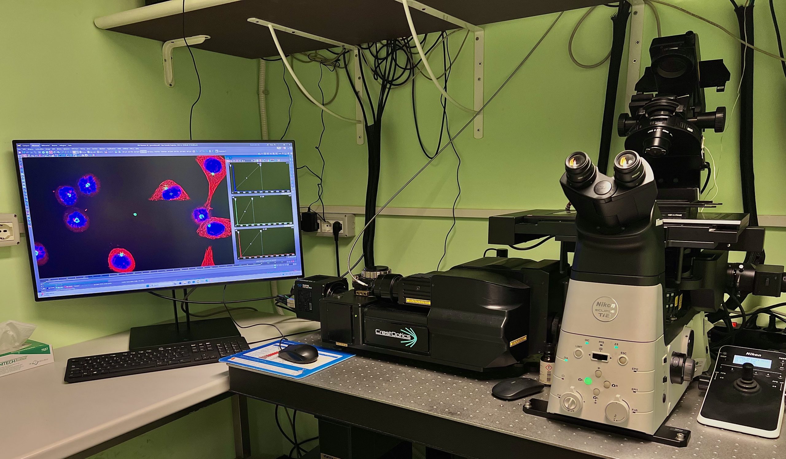
Microscope set-up located in the IPBM-CNR Imaging Platform in Rome.
“The IBPM Imaging Platform features the X-Light V3 Spinning Disk, offering the widest field of view currently available (25 mm). This flexible setup, supported by integrated environmental controls, enables optimal long-term live imaging over several days.”
Research Themes and Imaging Applications
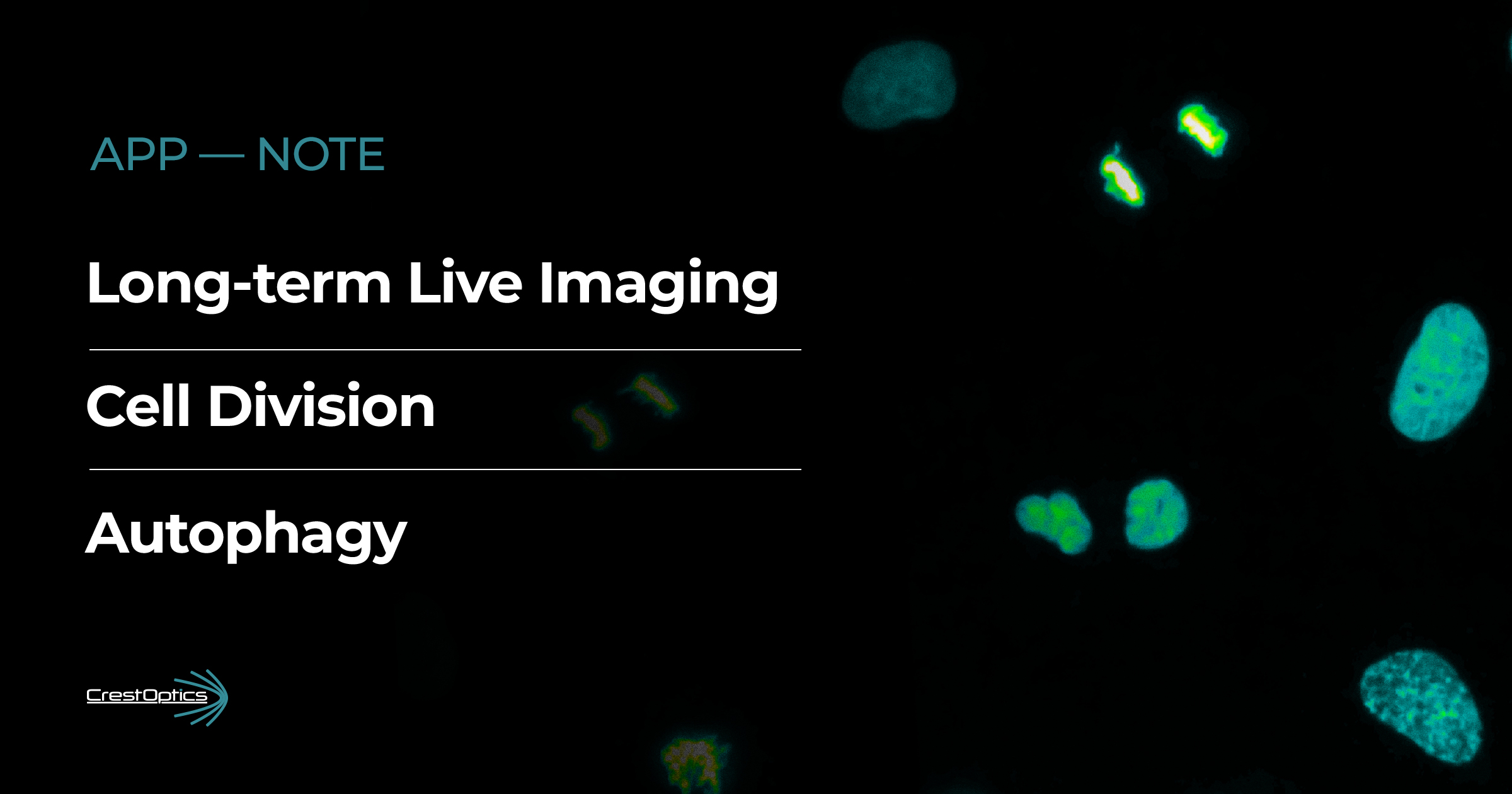
The research themes of IBPM groups who take advantage of the Imaging Platform are mainly focused on cell cycle and division, cell death and autophagy, cell migration, cellular differentiation, in both physiological and pathological conditions (e.g., cancer, neurological disorders), with the final aim of identifying novel therapeutic strategies. In this respect, the imaging platform offers unique possibilities in terms of real-time visualization of dynamic processes, visualization of cell heterogeneity and rare behaviors within a cell population, recording of cellular morphological changes in response to particular stimuli, high-definition analysis of subcellular structures and protein interactions (Figure 1). An interesting field of application of the spinning disk confocal microscope is the observation of advanced 3D cellular models, both in fixed samples and live dynamic assays. In recent years we have also dedicated our efforts to the development and use of high-content automated imaging pipelines, that are becoming essential to analyze the large image datasets generated with advanced imaging instrumentations. This requires collaborative and multidisciplinary interaction with colleagues from the mathematics field and with specialists from microscopy companies and yields useful tools that can be shared with the scientific community.
Figure 1: 19 hours time-lapse of U2OS cells stably expressing H2B-GFP. Frame interval 10 minutes, maximum Intensity Projection of a 7 μm z-stack. This image was acquired with the X-Light V3 with a 40x air objective (NA 0.95).
“The X-Light V3’s high-speed, low-phototoxic imaging allows us to visualize dynamic processes and advanced 3D models in real time. This provides unique insights into complex mechanisms which are essential for identifying novel therapeutic strategies.”
Case Studies that have also been described in the recent Application Note:
a) Mitosis
In our lab we have been investigating for several years the regulation of mitosis in mammalian cells. It is a key process that ensures balanced chromosome segregation in daughter cells (Figure 2). When altered it can lead to chromosomal instability and aneuploidy which are features of cancer cells; on the other hand, targeting mitosis is an effective strategy for anti-cancer therapy. Mitosis is highly dynamic and is characterized by spectacular morphological changes, therefore time-lapse microscopy is a highly informative approach to investigate it. At the same time, mitotic division is a highly sensitive process that can be altered or impaired by strong illumination under the microscope. Using the X-Light V3 microscope described before we have been able to record the mitotic process with multiple fluorescent markers and DIC, and high time resolution, enabling accurate measuring of mitotic entry, duration and aberrant phenotypes.By this time-lapse recording set-up we have recently revealed an unexpected role for a regulator of mitotic spindle orientation in an unexpected cell cycle window, i.e. before mitotic entry, thus unveiling a novel function that could not have been appreciated with other approaches.
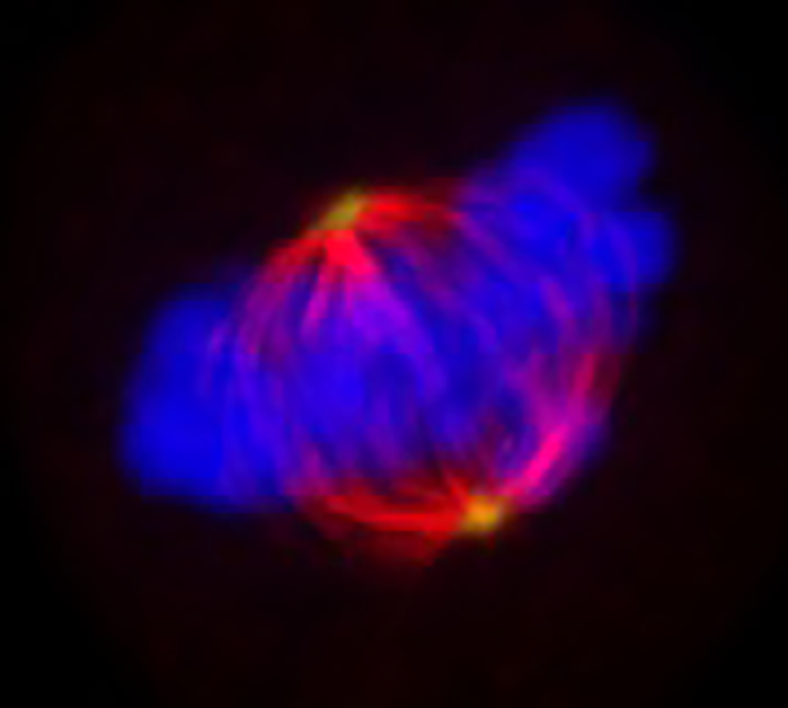
Figure 2: Composite immunofluorescence of a metaphase HeLa cell. Colors indicate alpha-tubulin (red), pericentrin (green), and DAPI-stained chromosomes (blue).
b) Authophagy
Research Focus: Neuroinflammation After Cardiopulmonary Bypass
Autophagy is a process where damaged protein aggregates or organelles are engulfed by a double-membrane vesicle called an autophagosome, which transports them to lysosomes for degradation and recycling (Figure 3). Given its role in maintaining cellular homeostasis, autophagy is essential for a variety of cellular processes. Our research in this field focuses on using pharmacological approaches to modulate autophagy in cancer cells to develop new therapeutic strategies. To understand how pharmacological compounds modulate specific stages of autophagy, it is necessary to visualise and quantitatively assess the formation, maturation and degradation of autophagosomes in real time. The X-Light V3 microscope, in conjunction with cellular tools expressing specific fluorescent markers (e.g. GFP-LC3 tandem probes and mRFP-GFP-LC3), facilitates the quantification of autophagosomes and autolysosomes, along with the assessment of autophagic flux (the rate of autophagosome formation and degradation), in both viable and fixed samples. This approach enabled detailed analysis of how novel pharmacological modulators affect distinct stages of autophagy, providing mechanistic insight into their mode of action. Additionally, the use of automated image analysis software ensures unbiased, statistically robust quantification across large cell populations.
Figure 3: 15 hours time-lapse of the H2199 mRFP-GFP-LC3 cell line. Frame interval 15 minutes. This image was acquired with the X-Light V3 with a 40x air objective (NA 0.95).
Overall, these case studies demonstrate the potential of spinning disk confocal microscopy with the X-Light V3, offering high-speed, low-phototoxic live-cell imaging. This provides valuable insights into complex processes such as cell division and autophagy, which are not readily discernible through alternative imaging methods.
Related Scientific Publications from the IBPM-CNR Imaging Platform Featuring Our Technology
Naso FD, Polverino F, Cilluffo D, Latini L, Stagni V, Asteriti IA, Rosa A, Soddu S, Guarguaglini G. Biochim Biophys Acta Mol Basis Dis. 2024 Apr;1870(4):167116. doi: 10.1016/j.bbadis.2024.167116. Epub 2024 Mar 4.PMID: 38447882
Pellegrini FR, De Martino S, Fianco G, Ventura I, Valente D, Fiore M, Trisciuoglio D, Degrassi F. Autophagy. 2023 Jul;19(7):2078-2093. doi: 10.1080/15548627.2023.2170962. Epub 2023 Feb 10.PMID: 36704963
Antonelli L, Polverino F, Albu A, Hada A, Asteriti IA, Degrassi F, Guarguaglini G, Maddalena L, Guarracino MR. Sci Data. 2023 Oct 4;10(1):677. doi: 10.1038/s41597-023-02540-1.PMID: 37794110
Explore major discoveries made with our systems on our Publications page!
CrestOptics & Nikon Italy: Strengthening a Long-Lasting Partnership
Giacomo Cozzi
Nikon Italy and CrestOptics have been collaborating for more than 10 years in the field of advanced imaging. Our partnership began with the development of the first X-Light V1 together with the Nikon ECLIPSE Ti-E, and over the years we have grown side by side in the spinning disk sector with their innovative systems such as X-Light V2, CICERO, and DeepSIM.
The combination of the X-Light V3 and the Nikon ECLIPSE Ti2-E has proven to be a winning solution for customers requiring a high-end, fast-acquisition platform for live-cell confocal imaging. Thanks to the NIS-Elements software, we can further enhance the potential of the spinning disk platform by integrating market‑leading products such as Kinetix (Teledyne Photometrics) and Celesta (Lumencor Inc.). The result is a system capable of acquiring high‑resolution confocal images at lightning speed while ensuring minimal phototoxicity.
This level of quality and versatility has been greatly appreciated by the major imaging facilities across Italy, and we are particularly proud to have provided these capabilities to the Imaging Platform of IBPM‑CNR, one of the key microscopy hubs in the central‑southern region.
This long‑standing collaboration continues to inspire new technological advances and reinforces our shared commitment to supporting the scientific community with ever more powerful and reliable imaging solutions.