
Publications
Under the spotlight
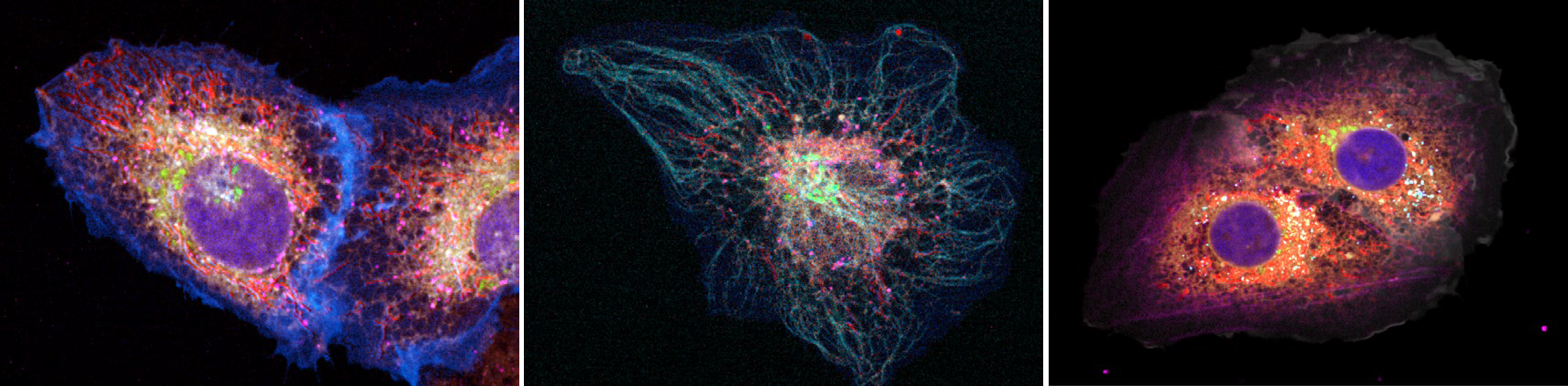
Multispectral live-cell imaging with uncompromised spatiotemporal resolution (Kumar et al., 2025; Nature Photonics)
Multispectral spinning-disk confocal acquisitions and Richardson-Lucy spectral unmixing (RLSU) results of live U2OS cells; (left) cell transfected with the six-color ColorfulCell polycistronic plasmid alone; (middle) ColorfulCell-transfected cell additionally incubated with SPY555-tubulin dye to visualize the microtubule network; (right) ColorfulCell-transfected cell additionally incubated with SPY555-actin dye to visualize the actin cytoskeleton. The images demonstrate accurate seven-color unmixing (nuclei, plasma membrane, mitochondria, Golgi, lysosomes/ER, peroxisomes, and the respective cytoskeletal dye) using a eight-channel camera-based hardware module. Images were acquired with the X-Light V3 and a custom-built multispectral acquisition module.
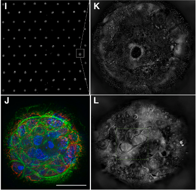
Mechanosensitive interactions of tumoroids with an engineered environment promote cell proliferation and enhance drug response detection (Ouni et al., 2025; Cell Biomaterials)
Representative images: (top and bottom left) high-content acquisitions of tumoroids in microarrays with confocal microscopy (green, F-actin; red, mitochondria; blue, nuclei); (top right) label-free holotomography revealing microvilli in interaction with the hydrogel; (bottom right) real-time monitoring of cell divisions and spindle formation with holotomography. Images were acquired with X-Light V2 and Tomocube’s HT-X1™ Plus.
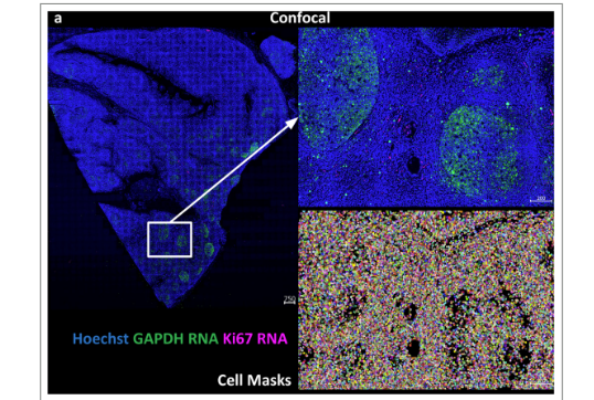
Modular, open-sourced multiplexing for democratizing spatial omics (Zhang et al., 2025; Lab on a Chip)
Representative confocal images from automated, RNA staining with Python-based robotic imaging and staining for modular spatial omics (PRISMS) on normal, adult human tonsil tissues. RNA labels stained GAPDH RNA (magenta) and Ki67 RNA (green). Hoechst is shown in blue. The bottom row illustrates the single-cell masks. Below, single-cell expression quantification from confocal imaging of RNA. Images were acquired with CICERO, integrated in a Cephla setup.
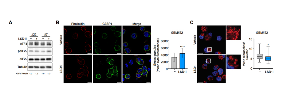
Metabolic traits shape responses to LSD1-directed therapy in glioblastoma tumor-initiating cells (Marotta et al., 2025; Science Advances)
Figure demonstrating that LSD1 inhibition (LSD1i) activates the integrated stress response (ISR) and induces early mitochondrial structural alterations in glioblastoma tumor-initiating cells (GBM TICs). Panel A shows increased levels of ISR markers by western blot, indicating stress pathway activation. Panel B reveals the formation of G3BP1-positive stress granules, further confirming a cellular stress response. Panel C shows that LSD1i leads to mitochondrial fragmentation, highlighting structural mitochondrial disruption as an early consequence of treatment. These images were acquired with DeepSIM (Figure B) and X-Light V3.
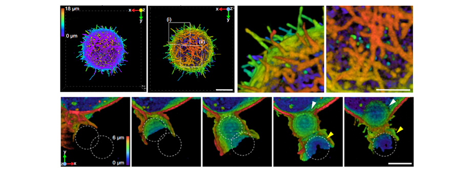
Unveiling cellular communications through rapid pan-membrane-protein labeling (Gunasekara et al., 2025; Nature Communications)
Super Resolution volumetric images showing that pan-membrane high-density labeling enables unprecedented visualization of: fine membrane structures (microvilli, ruffles) and dynamic processes like phagocytosis and interfacial material transfer. Images were acquired with DeepSIM.
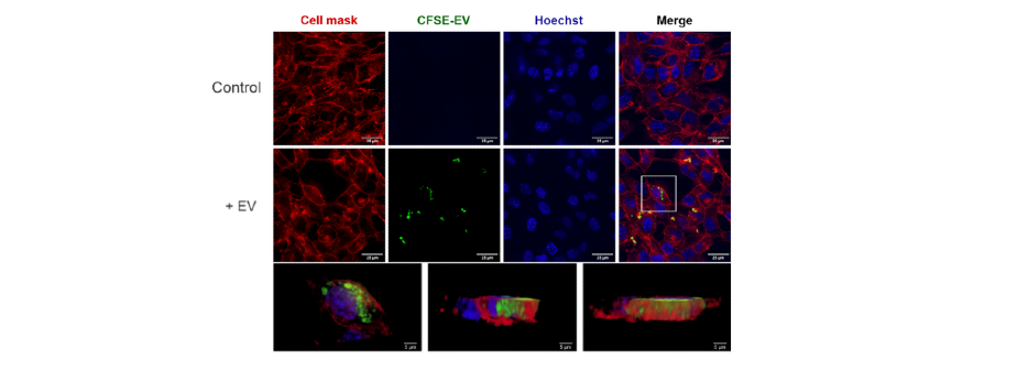
CCR2 cooperativity promotes hematopoietic stem cell homing to the bone marrow (Hurwitz et al., 2024; Science Advances)
Confocal microscopy images of Hematopoietic Stem and Progenitor Cells (HSPC) -derived extracellular vescicles (EVs) uptake by bone marrow endothelial cells. Control cells show no green fluorescence, indicating no EVs uptake. In the +EV condition, carboxyfluorescein succinimidyl ester (CFSE) -labeled EVs (green) colocalize with red membrane markers, confirming internalization. 3D views further demonstrate intracellular localization. Images were acquired with X-Light V3.
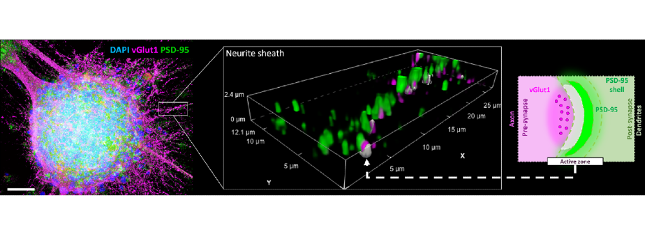
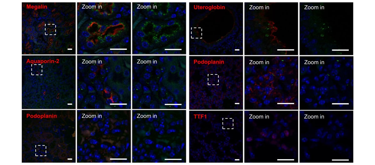
Extracellular PHF‑tau modulates astrocyte mitochondrial dynamics and mediates neuronal connectivity. (Zufferey et al., 2025; Translational Neurodegeneration)
Combined illustration and methodological overview of neurospheroid immunostaining analysis. Left: confocal image of a neurospheroid stained for Vglut and PSD-95 to study the effect of Tau protein on synaptic and neuronal balance. Scale bar: 100 μm. Center: inset showing an example of a 3D volume of interest analyzed for synaptic markers. Right: schematic illustrating the approach used for segmenting active zones and postsynaptic compartments. Images were acquired with
X-Light V3.
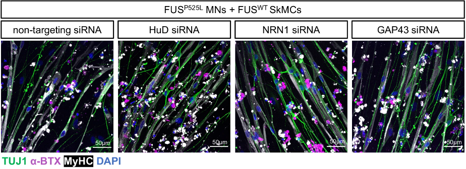
HuD impairs neuromuscular junctions and induces apoptosis in human iPSC and Drosophila ALS models (Silvestri et al., 2024; Nature Communications)
Confocal imaging of co-cultures of FUSP525L motoneurons and FUS wild-type skeletal muscle cells, treated with siRNAs targeting HuD, NRN1, and GAP43. Images were acquired with X-Light V3.
A prenatal skin atlas reveals immune regulation of human skin morphogenesis (Gopee et al.; 2024; Nature)
Confocal imaging of the skin organoid model illustrating the interaction between immune and non-immune cells in the development of the vascular network in prenatal skin. Images were acquired with X-Light V3.
Transport and Organization of Individual Vimentin Filaments Within Dense Networks Revealed by Single Particle Tracking and 3D FIB-SEM (Renganathan et al., 2024; BioRxiv)
Super Resolution image of a fixed Human Retinal Pigment Epithelial (RPE) cell expressing Vimentin-SunTag (green) and immunolabeled for endogenous vimentin (magenta). This image was acquired with DeepSIM.
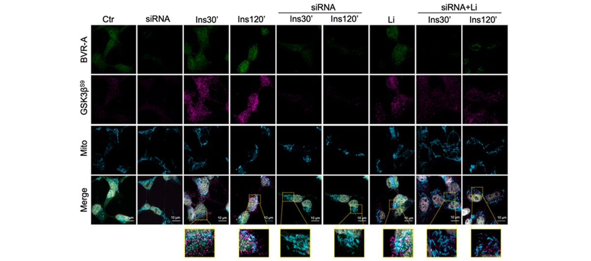
Biliverdin Reductase-A integrates insulin signaling with mitochondrial metabolism through phosphorylation of GSK3β (Lanzillotta et al., 2024; Redox Biology)
Representative confocal images of Biliverdin Reductase-A (green), Mitotracker (cyan) and GSK3βS9 (magenta) in Ctr and siRNA-treated cells stimulated with 100 nM insulin. Images were acquired with X-Light V3.
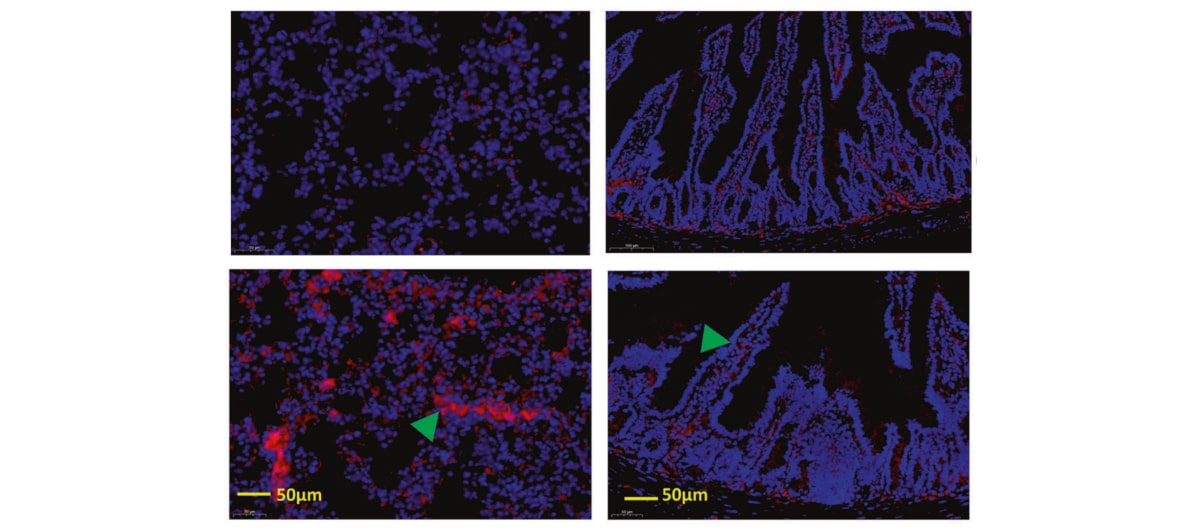
Lactate exacerbates lung damage induced by nanomicroplastic through the gut microbiota–HIF1a/PTBP1 pathway (Xuan et al., 2023 Experimental & molecular medicine)
In this figure is shown the increase of N-cadherin in the lung and intestinal tissues of mice treated with nano-MPs, indicating that nano-MPs can simultaneously damage the lungs and intestines. This image was acquired with X-Light V3.
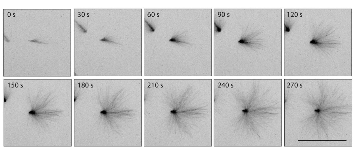
Branched microtubule nucleation and dynein transport organize RanGTP asters in Xenopus laevis egg extract (Scrofani et al., 2023 Molecular biology of the cell)
This figure represents a time lapse of dynamic MT reassembling after temporally controlled perturbations of nucleation and dynein activity, demonstrating MT self-organizing activity. This image was acquired with X-Light V2.
Exosomes define a local and systemic communication network in healthy pancreas and pancreatic ductal adenocarcinoma (Adem et al., 2024 Nature Communications)
Representative confocal microscopy images of PDAC CD63+ exososomes accumulation in the kidneys or lungs of mice at a early Pancreatic Ductal Adenocarcinoma stages. This image was acquired with X-Light V3.
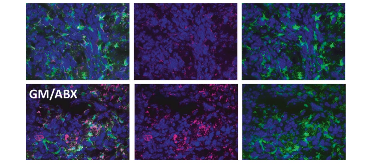
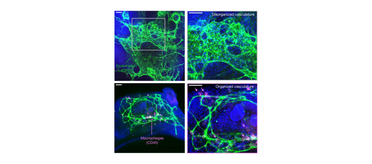
Antibiotics treatment promotes vasculogenesis in the brain of glioma-bearing mice (Rosito et al., 2024 Cell death and disease)
Images acquired with X-Light V3, showing representative z-projection images of microglia / macrophages cells in the tumoral area from glioma (GM) and glioma/antibiotics (GM/ABX) mice.

The symbol denotes articles published in journals with an impact factor of 10 or higher.
Sorted Publications based on first author affiliation country
Sorted publications based on keyword
Explore our product page and uncover our cutting-edge microscopy instruments that facilitate revolutionary breakthroughs across various research fields