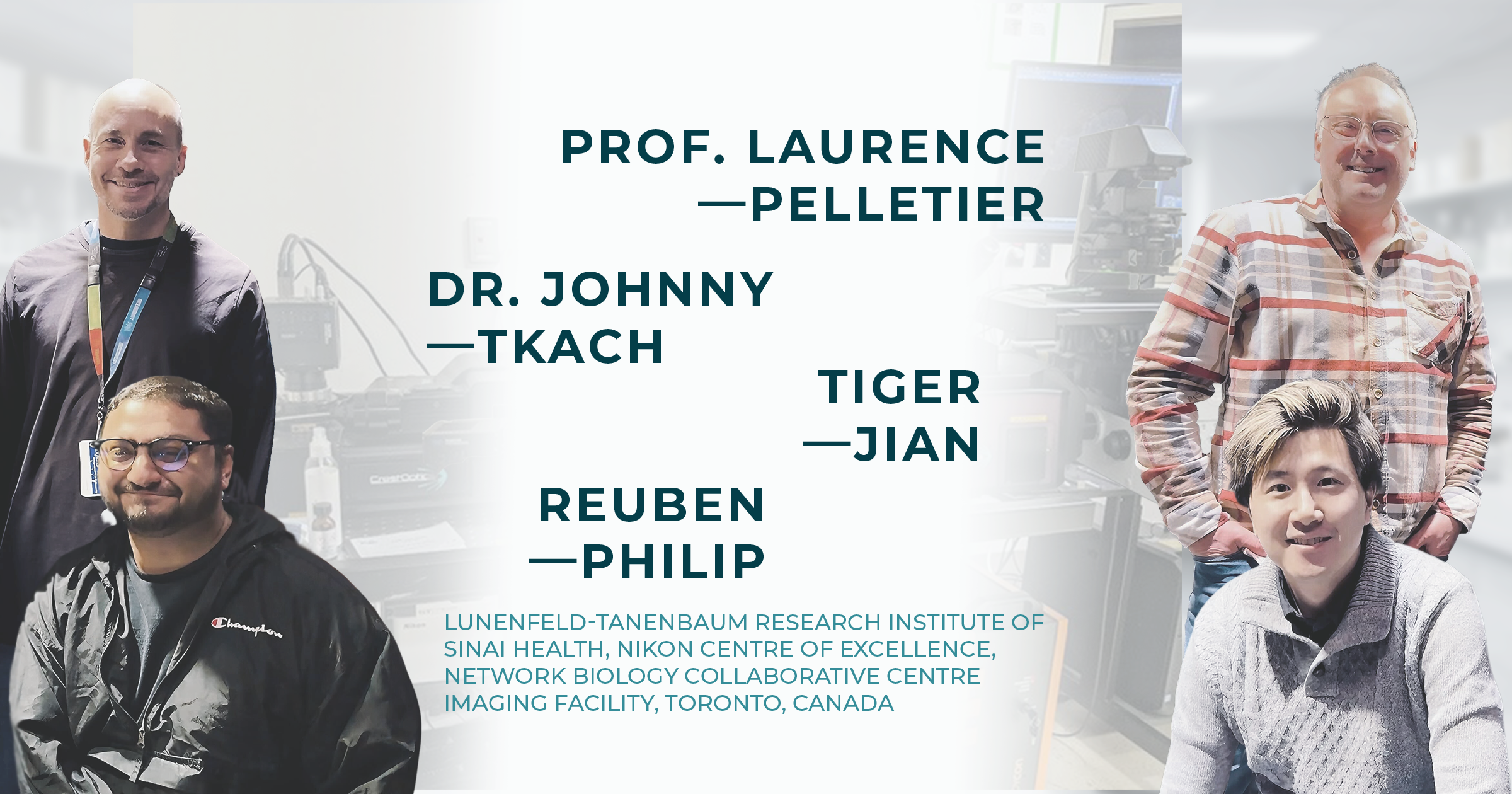
Interview with Prof. Laurence Pelletier (Senior Investigator / Director of the Nikon CoE a LTRI / Co-director a the NBCC), Dr. Johnny Tkach (Scientific Associate), Tiger Jian (Ph.D. Candidate) and Reuben Philip (Ph.D. Candidate)
Lunenfeld-Tanenbaum Research Institute (LTRI) of Sinai Health, Nikon Centre of Excellence (CoE), Network Biology Collaborative Centre (NBCC) Imaging Facility, Toronto, Canada
Introduction of the Nikon Centre of Excellence in Toronto
Located in the Lunenfeld-Tanenbaum Research Institute of Sinai Health, in the heart of Downtown Toronto, this first Canadian Nikon Centre of Excellence seeks to provide access to the latest in imaging technologies to its trainees, visiting scholars, and collaborators at the University of Toronto, the greater Toronto community and around the world. The goal of this Nikon Centre of Excellence is to harness the power of advanced imaging and image analysis to address the most salient questions in cell biology and to foster meaningful relationships between researchers and Nikon’s hardware and software development teams.Research Themes in the Pelletier Lab
The Pelletier Lab is fundamentally driven by a fascination with the construction and function of cellular machines, specifically focusing on how these organelles, whether membrane-bound like the cilia or non-membrane-bound like the centrosome, regulate complex cellular processes. Their research program centers on the dramatic morphological and functional transformations these structures undergo in tight coordination with the cell cycle. At the molecular level, the lab investigates the mechanisms governing centrosome- and cilia-related activities, ranging from centriole duplication and pericentriolar material assembly to ciliogenesis, cell motility, and mitotic spindle formation.
To decode these processes, the team utilizes a sophisticated toolkit of quantitative imaging, where they rely heavily on spinning disk confocal technology to achieve high-speed, live-cell visualization with minimal phototoxicity. This is integrated with a spectrum of approaches from standard to super-resolution microscopy, allowing for high-content screening and advanced morphometric measurements in systems ranging from single cells to developing organoids.
This imaging prowess is integrated with functional proteomics to systematically map the protein-protein interactions essential for centrosome and cilia function. By linking these proteomic datasets with genome-scale RNAi and CRISPR/Cas9 screens, the Pelletier Lab seeks to understand how these molecular pathways are disrupted in cancer and clinically relevant disorders such as microcephaly and various ciliopathies, ultimately aiming to define how their proper function ensures normal tissue homeostasis.
“We rely heavily on spinning disk confocal technology to achieve high-speed, live-cell visualization with minimal phototoxicity. This allows us to perform high-content screening and advanced morphometric measurements in systems ranging from single cells to developing organoids.”
Microscope Configuration: Setup Integrating the X-Light V3 & DeepSIM System
The lab utilizes a high-performance imaging pipeline centered on the CrestOptics X-Light V3 & DeepSIM X-Light system, integrated into a Nikon Ti2 Inverted microscope platform.
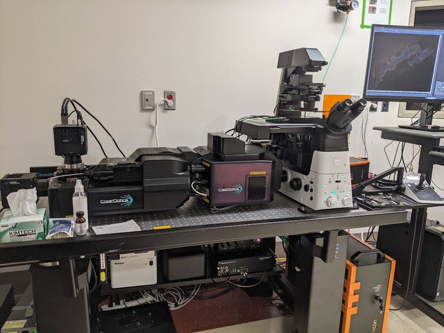
Microscope set-up at the Pelletier Lab, Nikon Center of Excellence in Toronto.
This setup is engineered for extreme sensitivity and speed, featuring a class-leading 25 mm field of view and dual back-illuminated Kinetix sCMOS cameras. A defining technical advantage of this configuration is its instantaneous modularity: with a single click, researchers can transition from widefield illumination to 7-line laser spinning disk confocal (ranging from violet to NIR) and into DeepSIM super-resolution mode. This allows the lab to resolve sub-diffraction structures, such as individual centriolar triplets, with unprecedented clarity.
To support the lab’s longitudinal live-cell assays, the platform maintains rigorous focal and environmental stability over multi-day experiments.
By employing the Nikon Perfect Focus System (PFS) alongside an Okolab stage incubator to regulate CO2, humidity, and temperature, the lab can track the complete centrosome duplication cycle with minimal phototoxic stress. All data acquisition and complex morphometric analyses are unified through Nikon NIS-Elements software.
“This setup is engineered for extreme sensitivity and speed, featuring a class-leading 25 mm field of view, allowing the lab to resolve sub-diffraction structures, such as individual centriolar triplets, with unprecedented clarity.”
Microscope Configuration: Setup Integrating the X-Light V3 & DeepSIM System
The lab utilizes a high-performance imaging pipeline centered on the CrestOptics X-Light V3 & DeepSIM X-Light system, integrated into a Nikon Ti2 Inverted microscope platform.
This setup is engineered for extreme sensitivity and speed, featuring a class-leading 25 mm field of view and dual back-illuminated Kinetix sCMOS cameras. A defining technical advantage of this configuration is its instantaneous modularity: with a single click, researchers can transition from widefield illumination to 7-line laser spinning disk confocal (ranging from violet to NIR) and into DeepSIM super-resolution mode. This allows the lab to resolve sub-diffraction structures, such as individual centriolar triplets, with unprecedented clarity.
To support the lab’s longitudinal live-cell assays, the platform maintains rigorous focal and environmental stability over multi-day experiments.
By employing the Nikon Perfect Focus System (PFS) alongside an Okolab stage incubator to regulate CO2, humidity, and temperature, the lab can track the complete centrosome duplication cycle with minimal phototoxic stress. All data acquisition and complex morphometric analyses are unified through Nikon NIS-Elements software.
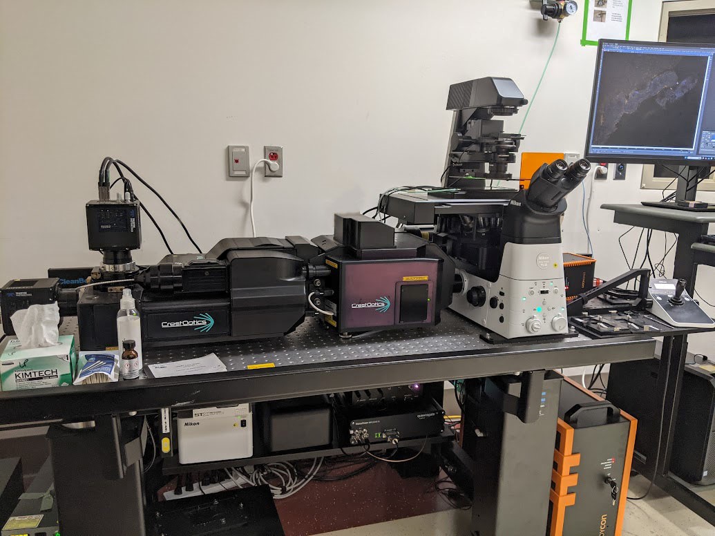
Microscope set-up at the Pelletier Lab, Nikon Center of Excellence in Toronto.
“This setup is engineered for extreme sensitivity and speed, featuring a class-leading 25 mm field of view, allowing the lab to resolve sub-diffraction structures, such as individual centriolar triplets, with unprecedented clarity.”
Case Studies
1) Dr. Johnny Tkach: Resolution for Neural Network Training in Mitotic Research
The flexibility of the X-Light V3 + DeepSIM microscope has been invaluable to our research. With a single system, we can perform high-resolution whole-organoid imaging, super-resolution microscopy, and high-speed dynamic 3D imaging. For every application, the large field of view enables us to capture more data per image, greatly improving our throughput and our ability to characterize phenotypes. We are currently using the system to investigate centrosome clustering during mitosis (Figure 1). The X-Light V3 allows us to image multiple points in 3D with exceptional temporal and spatial resolution, data that we use to train convolutional neural network models to assess mitotic stages and outcomes.
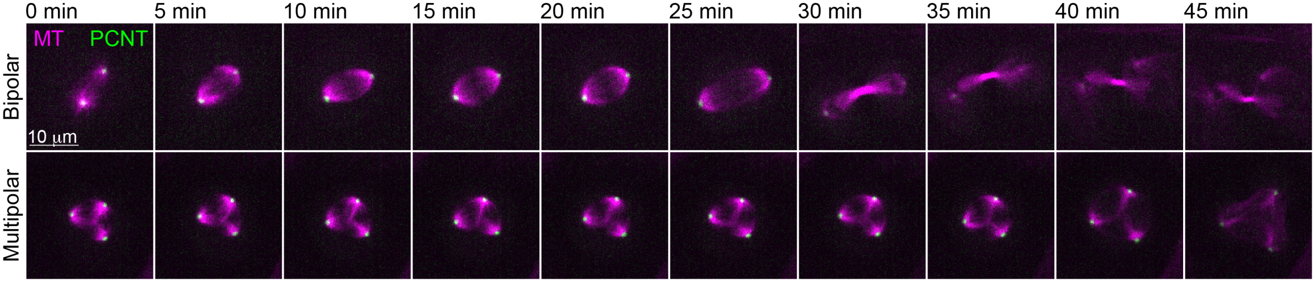
Figure 1: RPE-1 cells expressing mStayGold-PCNT (centrosome) and stained with SiR-tubulin (microtubules) were imaged every 5 min for 18h in 3D. Examples of maximum intensity projections of a normal bipolar mitotic event and an abnormal multipolar division are shown. Scale bar: 10 µm.
"Compared to other systems, the X-Light V3 spinning disk is exceptionally fast, making it highly effective for volumetric imaging of large 3D tissues. By utilizing its fast time-lapse capabilities, we achieve the crucial temporal resolution needed for highly dynamic functional assays, all while maintaining an excellent field of view."
2) Tiger Jian: Large Scale 3D Imaging and Temporal Resolution
The X-Light V3 spinning disk system is highly optimized for applications that demand exceptional imaging speed over expansive volumes, such as capturing large 3D tissues and organoids (Figure 2). For these macro-scale samples, where efficiently covering extensive XY and Z ranges is more critical than the extreme sub-cellular resolution of slower line-scanning confocals, the X-Light V3 paired with a Piezo stage delivers outstanding performance. Additionally, the system’s ultra-fast acquisition capabilities are instrumental for highly dynamic functional assays, such as tracking vascular perfusion with fluorescent beads. By utilizing “fast time-lapse” settings, the X-Light V3 can achieve impressive speeds of up to 500 frames per second (FPS), providing the crucial temporal resolution needed to make these rapid assays viable while maintaining an excellent field of view.
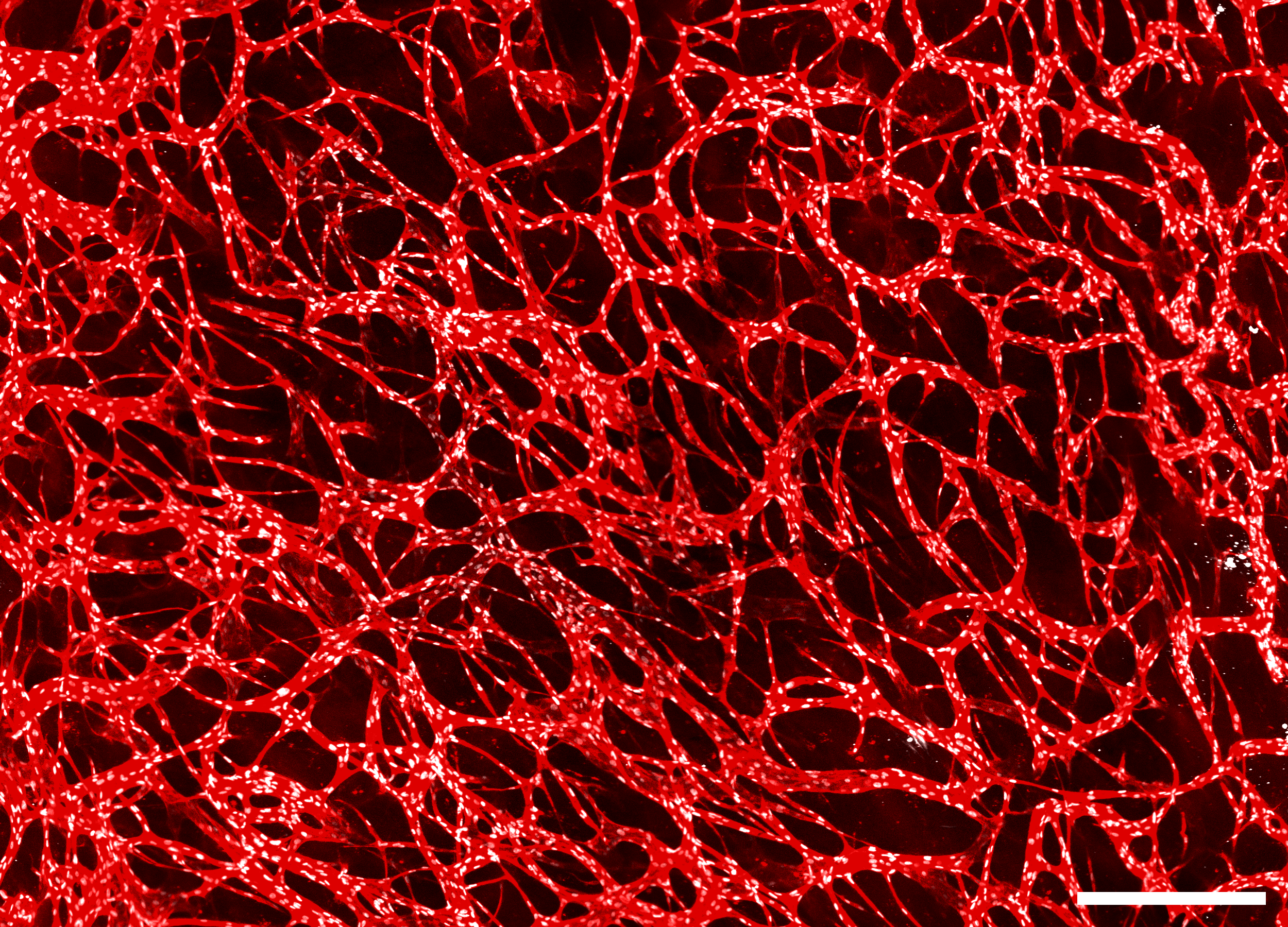
Figure 2: In vitro vascular network imaged on the X-Light V3 system. Red: vessels (CD31). White: endothelial nuclei (ERG). Scale bar: 500 µm.
“I find the X-Light V3 spinning disk to be exceptionally fast, making it highly effective for volumetric imaging of large 3D tissues. Additionally, the system's rapid frame rates are instrumental to the performance of our dynamic functional assays.”
3) Reuben Philip: Seamless Speed and Super-Resolution in One Platform
The combination of the X-Light V3 and DeepSIM offers researchers a powerful and user-friendly platform for both super-resolution and high-speed imaging. For fixed samples, DeepSIM paired with a 60X objective provides excellent resolution while preserving a broad field of view, allowing multiple cells to be imaged at once, an important advantage over other super-resolution systems with more limited imaging areas (Figure 3). For live-cell applications, the remarkable speed of the X-Light V3 enables the capture of rapid dynamic events such as EB1 comets. In addition, the system’s seamless dual-camera integration with Nikon’s NIS-Elements software removes the need for manual filter switching, making the workflow especially intuitive compared to other multimodal platforms that combine spinning disk and super-resolution imaging.
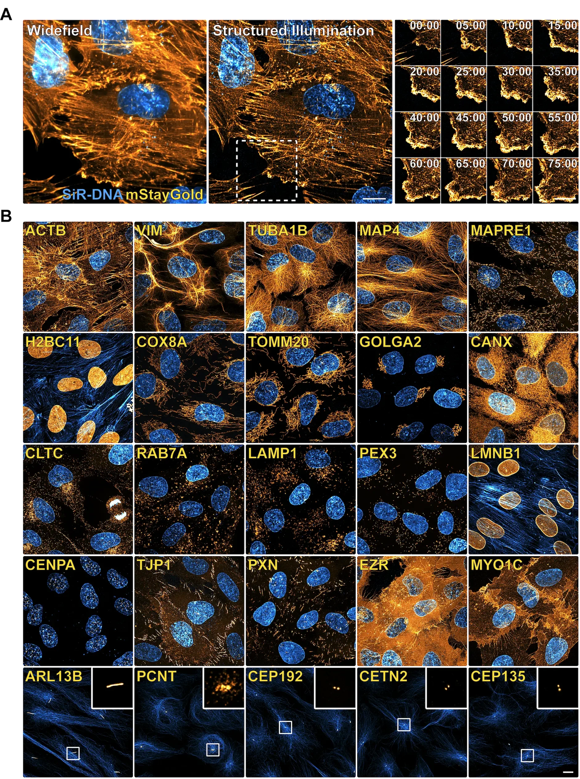
Figure 3: Endogenous mStayGold fusions generated by CRISPR using optimized qTAG donors. (A) Super-resolution timelapse imaging of mStayGold tagged ACTB (Orange) co-stained with SiR-DNA (Blue). (A – Left) – A widefield image at the start of the timelapse. (A – Middle) – The corresponding structured illumination super-resolution reconstructed image. (A – Right) – A time-lapse array of images following the highlighted area. (B) Shown are representative images of 25 genetically edited genes harbouring the mStayGold fluorescent protein sequence (Orange), co-stained with live SiR dyes (Blue) against Actin, Tubulin, or DNA, enabling visualization of their native expression, localization, and dynamics. Imaged via live DeepSIM. Scale bar: 10 µm. (Adapted from Philip et al., 2025. EMBO Journal).
“We have had great success using the X-Light V3 to capture rapid, dynamic live-cell phenomena such as EB1 comets.”
Related Scientific Publications from the Pelletier Lab Featuring Our Technology
(i) qTAG: an adaptable plasmid scaffold for CRISPR-based endogenous tagging.
Philip, R, Sharma, A, Matellan, L, Erpf, AC, Hsu, WH, Tkach, JM, Wyatt, HDM., Pelletier, L. The EMBO Journal, 2025; 44(3): 947-974. doi: 10.1038/s44318-024-00337-5.
(ii) A fast blind zero-shot denoiser.
Lequyer, J, Philip, R, Sharma, A, Hsu, WH, Pelletier, L. Nature Machine Intelligence, 2022; 4(11):953-963. doi: 10.1038/s42256-022-00547-8.
Jian, THZ, Sivitilli, AA, Guo, YE, Stirton, CJ, Gosio, JT, Tsukahara, Y, Tkach, JM, Lu, S, Yarmand, A, Mangos, M, Bremner, R, Wrana, JL, Attisano, L, Pelletier, L. Submitted Manuscript, 2026.
Explore major discoveries made with our systems on our Publications page!
CrestOptics & Nikon Canada Inc.
CrestOptics would like to extend its sincere thanks to Nikon Canada Inc for their ongoing support. This collaboration is a proud part of the Nikon Centre of Excellence, and we look forward to our continued partnership in bringing advanced CrestOptics imaging solutions to researchers across the country.